Chiasmal syndrome
Chiasmal syndrome refers to a collection of signs and symptoms that arise due to lesions affecting the optic chiasm. The optic chiasm is a critical structure located at the base of the brain where the optic nerves partially cross. This crossing allows for the visual information from the nasal (inner) half of each retina to be processed by the opposite side of the brain, which is essential for binocular vision.
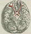
Anatomy of the Optic Chiasm[edit]
The optic chiasm is situated just above the pituitary gland and below the hypothalamus. It is a crucial part of the visual pathway, where the optic nerves from each eye converge and partially decussate. This anatomical arrangement is vital for the integration of visual information from both eyes, allowing for depth perception and a wide field of view.
Causes of Chiasmal Syndrome[edit]
Chiasmal syndrome can be caused by various conditions that affect the optic chiasm, including:
- Pituitary adenomas: These are the most common cause of chiasmal syndrome. As these tumors grow, they can compress the optic chiasm, leading to visual disturbances.
- Craniopharyngiomas: These are benign tumors that can also exert pressure on the optic chiasm.
- Meningiomas: Tumors arising from the meninges can affect the optic chiasm if they occur in the parasellar region.
- Aneurysms: Vascular abnormalities such as aneurysms of the internal carotid artery can impinge on the optic chiasm.
- Trauma: Head injuries can sometimes lead to damage to the optic chiasm.
Symptoms of Chiasmal Syndrome[edit]
The hallmark symptom of chiasmal syndrome is a specific type of visual field defect known as bitemporal hemianopsia. This occurs because the crossing fibers from the nasal retinae are affected, leading to loss of vision in the outer (temporal) fields of both eyes. Other symptoms may include:
- Decreased visual acuity
- Dyschromatopsia (color vision defects)
- Optic atrophy
- Headaches
Diagnosis[edit]
Diagnosis of chiasmal syndrome typically involves a combination of clinical evaluation and imaging studies. Magnetic resonance imaging (MRI) is the preferred modality for visualizing the optic chiasm and identifying any lesions. Visual field testing is also crucial to assess the extent and pattern of visual field loss.
Treatment[edit]
Treatment of chiasmal syndrome depends on the underlying cause. Options may include:
- Surgical resection of tumors
- Radiation therapy
- Medical management of pituitary adenomas with medications such as dopamine agonists
Prognosis[edit]
The prognosis for patients with chiasmal syndrome varies depending on the cause and the timeliness of treatment. Early intervention can often lead to significant improvement in visual function, especially if the underlying cause is a treatable tumor.
Related Pages[edit]
-
 Chiasmal syndrome


Medical Disclaimer: WikiMD is for informational purposes only and is not a substitute for professional medical advice. Content may be inaccurate or outdated and should not be used for diagnosis or treatment. Always consult your healthcare provider for medical decisions. Verify information with trusted sources such as CDC.gov and NIH.gov. By using this site, you agree that WikiMD is not liable for any outcomes related to its content. See full disclaimer.
Credits:Most images are courtesy of Wikimedia commons, and templates, categories Wikipedia, licensed under CC BY SA or similar.
Translate page: - East Asian
中文,
日本,
한국어,
South Asian
हिन्दी,
தமிழ்,
తెలుగు,
Urdu,
ಕನ್ನಡ,
Southeast Asian
Indonesian,
Vietnamese,
Thai,
မြန်မာဘာသာ,
বাংলা
European
español,
Deutsch,
français,
Greek,
português do Brasil,
polski,
română,
русский,
Nederlands,
norsk,
svenska,
suomi,
Italian
Middle Eastern & African
عربى,
Turkish,
Persian,
Hebrew,
Afrikaans,
isiZulu,
Kiswahili,
Other
Bulgarian,
Hungarian,
Czech,
Swedish,
മലയാളം,
मराठी,
ਪੰਜਾਬੀ,
ગુજરાતી,
Portuguese,
Ukrainian
