The Patella
Anatomy > Gray's Anatomy of the Human Body > II. [Osteology]] > 6c. 4. The Patella
Henry Gray (1821–1865). Anatomy of the Human Body. 1918.
The Patella[edit]
(Knee Cap)
The patella (Figs. 255, 256) is a flat, triangular bone, situated on the front of the knee-joint. It is usually regarded as a sesamoid bone, developed in the tendon of the Quadriceps femoris, and resembles these bones (1) in being developed in a tendon; (2) in its center of ossification presenting a knotty or tuberculated outline; (3) in being composed mainly of dense cancellous tissue. It serves to protect the front of the joint, and increases the leverage of the Quadriceps femoris by making it act at a greater angle. It has an anterior and a posterior surface three borders, and an apex.


Surfaces[edit]
The anterior surface is convex, perforated by small apertures for the passage of nutrient vessels, and marked by numerous rough, longitudinal striae. This surface is covered, in the recent state, by an expansion from the tendon of the Quadriceps femoris, which is continuous below with the superficial fibers of the ligamentum patellae. It is separated from the integument by a bursa.
The posterior surface presents above a smooth, oval, articular area, divided into two facets by a vertical ridge; the ridge corresponds to the groove on the patellar surface of the femur, and the facets to the medial and lateral parts of the same surface; the lateral facet is the broader and deeper. Below the articular surface is a rough, convex, non-articular area, the lower half of which gives attachment to the ligamentum patellae; the upper half is separated from the head of the tibia by adipose tissue.
Borders[edit]
The base or superior border is thick, and sloped from behind, downward, and forward: it gives attachment to that portion of the Quadriceps femoris which is derived from the Rectus femoris and Vastus intermedius.
The medial and lateral borders are thinner and converge below: they give attachment to those portions of the Quadriceps femoris which are derived from the Vasti lateralis and medialis.
Apex[edit]
The apex is pointed, and gives attachment to the ligamentum patellae.
Structure[edit]
The patella consists of a nearly uniform dense cancellous tissue, covered by a thin compact lamina. The cancelli immediately beneath the anterior surface are arranged parallel with it. In the rest of the bone they radiate from the articular surface toward the other parts of the bone.
Ossification[edit]
The patella is ossified from a single center, which usually makes its appearance in the second or third year, but may be delayed until the sixth year. More rarely, the bone is developed by two centers, placed side by side. Ossification is completed about the age of puberty.
Articulation[edit]
The patella articulates with the femur.
Function[edit]
The primary functional role of the patella is knee extension. The patella increases the leverage that the quadriceps tendon can exert on the femur by increasing the angle at which it acts.
The patella is attached to the tendon of the quadriceps femoris muscle, which contracts to extend/straighten the knee. The patella is stabilized by the insertion of the horizontal fibres of vastus medialis and by the prominence of the lateral femoral condyle, which discourages lateral dislocation during flexion. The retinacular fibres of the patella also stabilize it during exercise.
Additional images[edit]
-
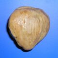 Human left patella from the front
 -
 Human left patella from behind
 -
 Flexion and extension of knee


External links[edit]
| Bones of the human leg | ||||||||||||||||||||||||||
|---|---|---|---|---|---|---|---|---|---|---|---|---|---|---|---|---|---|---|---|---|---|---|---|---|---|---|
|
Gray's Anatomy[edit]
- Gray's Anatomy Contents
- Gray's Anatomy Subject Index
- About Classic Gray's Anatomy
- Glossary of anatomy terms
Anatomy atlases (external)[edit]
[1] - Anatomy Atlases
| Human systems and organs | ||||||||||||||
|---|---|---|---|---|---|---|---|---|---|---|---|---|---|---|
|

Medical Disclaimer: WikiMD is for informational purposes only and is not a substitute for professional medical advice. Content may be inaccurate or outdated and should not be used for diagnosis or treatment. Always consult your healthcare provider for medical decisions. Verify information with trusted sources such as CDC.gov and NIH.gov. By using this site, you agree that WikiMD is not liable for any outcomes related to its content. See full disclaimer.
Credits:Most images are courtesy of Wikimedia commons, and templates, categories Wikipedia, licensed under CC BY SA or similar.
Translate page: - East Asian
中文,
日本,
한국어,
South Asian
हिन्दी,
தமிழ்,
తెలుగు,
Urdu,
ಕನ್ನಡ,
Southeast Asian
Indonesian,
Vietnamese,
Thai,
မြန်မာဘာသာ,
বাংলা
European
español,
Deutsch,
français,
Greek,
português do Brasil,
polski,
română,
русский,
Nederlands,
norsk,
svenska,
suomi,
Italian
Middle Eastern & African
عربى,
Turkish,
Persian,
Hebrew,
Afrikaans,
isiZulu,
Kiswahili,
Other
Bulgarian,
Hungarian,
Czech,
Swedish,
മലയാളം,
मराठी,
ਪੰਜਾਬੀ,
ગુજરાતી,
Portuguese,
Ukrainian
