Orbit (anatomy): Difference between revisions
CSV import Tags: mobile edit mobile web edit |
CSV import |
||
| Line 42: | Line 42: | ||
{{Skull}} | {{Skull}} | ||
{{stub}} | {{stub}} | ||
<gallery> | |||
File:Eye_orbit_anatomy_anterior2.jpg|Anterior view of the eye orbit anatomy | |||
File:Orbit_of_the_Face,_STL_file_processed_1.0.0.stl|3D model of the orbit of the face | |||
File:Orbital_bones.png|Diagram of the orbital bones | |||
File:Tear_system.svg|Diagram of the tear system | |||
File:Orbita2.jpg|Orbit (anatomy) | |||
File:Gray192.png|Anatomy of the orbit from Gray's Anatomy | |||
File:Gray787.png|Muscles of the orbit from Gray's Anatomy | |||
File:Orbita_mensch.jpg|Orbit (anatomy) | |||
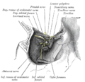
File:Lateral_orbit_nerves.jpg|Lateral view of the orbit showing nerves | |||
File:Temporal_fossa.jpg|Temporal fossa related to the orbit | |||
</gallery> | |||
Latest revision as of 11:29, 18 February 2025
Orbit (anatomy)
The Orbit is the cavity or socket of the skull in which the eye and its appendages are situated. The orbit is formed by the cheekbone, the forehead, the temple, and the side of the nose. The eye is cushioned within the orbit by pads of fat.
Structure[edit]
The orbit has a volume of 30 millilitres, and a height, width, and depth of approximately 40, 35, and 45 millimetres respectively. The entrance to the orbital cavity is referred to as the base, and the back as the apex. Four bones contribute to the formation of the orbital cavity.
Bones[edit]
The bones of the orbit include:
- The frontal bone
- The zygomatic bone
- The ethmoid bone
- The sphenoid bone
- The maxillary bone
- The palatine bone
- The lacrimal bone
Function[edit]
The primary function of the orbit is to provide a cavity in which the eye and its appendages are housed. This protects the eye from injury and allows for the movement of the eye.
Clinical significance[edit]
Conditions that can affect the orbit and therefore the eye include Orbital cellulitis, Orbital fracture, and Graves' disease.
See also[edit]
References[edit]
<references />
| Anatomy of the globe of the human eye | ||||||||||||||||||
|---|---|---|---|---|---|---|---|---|---|---|---|---|---|---|---|---|---|---|
|
| Neurocranium of the skull | ||||||||||||||||||||||||||||||||
|---|---|---|---|---|---|---|---|---|---|---|---|---|---|---|---|---|---|---|---|---|---|---|---|---|---|---|---|---|---|---|---|---|
|
 | |
|---|---|
|
|
 |
-
 Anterior view of the eye orbit anatomy
 -
 3D model of the orbit of the face
 -
 Diagram of the orbital bones
 -
 Diagram of the tear system
 -
 Orbit (anatomy)
 -
 Anatomy of the orbit from Gray's Anatomy
 -
 Muscles of the orbit from Gray's Anatomy
 -
 Orbit (anatomy)
 -
 Lateral view of the orbit showing nerves
 -
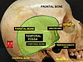 Temporal fossa related to the orbit


