Koniocellular cell
Koniocellular cells are a type of neuron found in the retina of the eye. They are part of the retinal ganglion cell layer and are involved in the processing of visual information. Koniocellular cells are smaller than other retinal ganglion cells and have a unique role in visual processing.
Anatomy[edit]
Koniocellular cells are located in the retinal ganglion cell layer of the retina. They are smaller than other retinal ganglion cells, such as parvocellular cells and magnocellular cells. Koniocellular cells have small, densely packed dendritic fields and are found throughout the retina.
Function[edit]
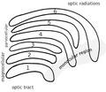
Koniocellular cells are involved in the processing of visual information. They receive input from bipolar cells and amacrine cells, and send output to the lateral geniculate nucleus (LGN) of the thalamus. Koniocellular cells are thought to play a role in color vision, contrast sensitivity, and spatial resolution.
Clinical significance[edit]
Abnormalities in koniocellular cells have been implicated in a number of visual disorders, including glaucoma, macular degeneration, and retinitis pigmentosa. Further research is needed to fully understand the role of koniocellular cells in these conditions.
See also[edit]
References[edit]

This WikiMD article can only be edited by registered and verified editors. You can log in or register.
-
 Koniocellular cell
 -
 Koniocellular cell


Medical Disclaimer: WikiMD is for informational purposes only and is not a substitute for professional medical advice. Content may be inaccurate or outdated and should not be used for diagnosis or treatment. Always consult your healthcare provider for medical decisions. Verify information with trusted sources such as CDC.gov and NIH.gov. By using this site, you agree that WikiMD is not liable for any outcomes related to its content. See full disclaimer.
Credits:Most images are courtesy of Wikimedia commons, and templates, categories Wikipedia, licensed under CC BY SA or similar.
Translate page: - East Asian
中文,
日本,
한국어,
South Asian
हिन्दी,
தமிழ்,
తెలుగు,
Urdu,
ಕನ್ನಡ,
Southeast Asian
Indonesian,
Vietnamese,
Thai,
မြန်မာဘာသာ,
বাংলা
European
español,
Deutsch,
français,
Greek,
português do Brasil,
polski,
română,
русский,
Nederlands,
norsk,
svenska,
suomi,
Italian
Middle Eastern & African
عربى,
Turkish,
Persian,
Hebrew,
Afrikaans,
isiZulu,
Kiswahili,
Other
Bulgarian,
Hungarian,
Czech,
Swedish,
മലയാളം,
मराठी,
ਪੰਜਾਬੀ,
ગુજરાતી,
Portuguese,
Ukrainian
