The Organ of Hearing: Difference between revisions
No edit summary Tag: visualeditor-wikitext |
No edit summary |
||
| Line 1: | Line 1: | ||
[[Anatomy]] > [[Gray's Anatomy| Gray's Anatomy of the Human Body]] > X. The Organs of the Senses and the Common Integument > 1d. The Organ of Hearing | |||
[[Henry Gray]] (1821–1865). Anatomy of the Human Body. 1918. | |||
== '''The Organ of Hearing''' == | |||
[[Henry Gray]] (1821–1865). | |||
== '''The Organ of Hearing''' | |||
'''(Organon Auditus; The Ear)''' | '''(Organon Auditus; The Ear)''' | ||
The '''[[ear]]''' or '''organ of hearing''' is divisible into three parts: | The '''[[ear]]''' or '''organ of hearing''' is divisible into three parts: | ||
The '''[[external ear]]''' | |||
The '''[[middle ear]]''' or '''[[tympanic cavity]]''' and | |||
The '''[[internal ear]]''' or '''[[labyrinth]]''' | |||
[[File:Gray898.png|thumb|FIG. 898– Section through the head of a human embryo, about twelve days old, in the region of the hind-brain. (Kollmann.) (''Picture From the Classic Gray's Anatomy'')]] | |||
[[File:Gray899.png|thumb|left|FIG. 899– Section through hind-brain and auditory vesicles of an embryo more advanced than that of Fig. 898. (After His.) (''Picture From the Classic Gray's Anatomy'')]] | |||
FIG. 900– Lateral views of membranous labyrinth and acoustic complex. X 25 dia. (Streeter.) ''[[absorpt. focu]]'' area of wall where absorption is complete; ''[[amp]]'' ampulla membranacea; ''[[crus]]'' crus commune; ''[[d. sc. lat]]'' ductus semicircularis lateralis; ''[[d. sc. post]]'' ductus semicircularis posterior; ''[[d. sc. sup]]'' ductus semicircular superior; ''[[coch. or cochlea]]'' ductus cochlearis; ''[[duct. endolymph]]'' ductus endolymphaticus; ''[[d. reuniens]]'' ductus reuniens Henseni; ''[[endol. or endolymphs]]'' appendix endolymphaticus; ''[[rec. utr]]'' recessus utriculi; ''[[sacc]]'' sacculus; ''[[sac. endol]]'' saccus endolymphaticus; ''[[sinus utr. lat]]'' sinus utriculi lateralis; ''[[utric]]'' utriculus; ''[[vestib. p]]'' vestibular pouch. (''Picture From the Classic Gray's Anatomy'') | [[File:Gray900.png|thumb|FIG. 900– Lateral views of membranous labyrinth and acoustic complex. X 25 dia. (Streeter.) ''[[absorpt. focu]]'' area of wall where absorption is complete; ''[[amp]]'' ampulla membranacea; ''[[crus]]'' crus commune; ''[[d. sc. lat]]'' ductus semicircularis lateralis; ''[[d. sc. post]]'' ductus semicircularis posterior; ''[[d. sc. sup]]'' ductus semicircular superior; ''[[coch. or cochlea]]'' ductus cochlearis; ''[[duct. endolymph]]'' ductus endolymphaticus; ''[[d. reuniens]]'' ductus reuniens Henseni; ''[[endol. or endolymphs]]'' appendix endolymphaticus; ''[[rec. utr]]'' recessus utriculi; ''[[sacc]]'' sacculus; ''[[sac. endol]]'' saccus endolymphaticus; ''[[sinus utr. lat]]'' sinus utriculi lateralis; ''[[utric]]'' utriculus; ''[[vestib. p]]'' vestibular pouch. (''Picture From the Classic Gray's Anatomy'')]] | ||
=== '''The Development of the Ear''' === | === '''The Development of the Ear''' === | ||
The first rudiment of the internal ear appears shortly after that of the eye, in the form of a patch of thickened [[ectoderm]], the '''[[auditory plate]]''' over the region of the [[hind-brain]]. The auditory plate becomes depressed and converted into the '''[[auditory pit]]''' (Fig. 898). The mouth of the pit is then closed, and thus a shut sac, the '''auditory vesicle''' is formed (Fig. 899); from it the epithelial lining of the membranous [[labyrinth]] is derived. The [[vesicle]] becomes pear-shaped, and the neck of the flask is obliterated (Fig. 900). From the vesicle certain [[diverticula]] are given off which form the various parts of the membranous labyrinth. | The first rudiment of the internal ear appears shortly after that of the eye, in the form of a patch of thickened [[ectoderm]], the '''[[auditory plate]]''' over the region of the [[hind-brain]]. The auditory plate becomes depressed and converted into the '''[[auditory pit]]''' (Fig. 898). The mouth of the pit is then closed, and thus a shut sac, the '''auditory vesicle''' is formed (Fig. 899); from it the epithelial lining of the membranous [[labyrinth]] is derived. The [[vesicle]] becomes pear-shaped, and the neck of the flask is obliterated (Fig. 900). From the vesicle certain [[diverticula]] are given off which form the various parts of the membranous labyrinth. | ||
One from the middle part forms the [[ductus]] and saccus endolymphaticus, another from the [[anterior]] end gradually elongates, and, forming a tube coiled on itself, becomes the [[cochlear duct]], the vestibular extremity of which is subsequently constricted to form the canalis reuniens. Three others appear as disk-like [[evaginations]] on the surface of the [[vesicle]]; the central parts of the walls of the disks [[coalesce]] and disappear, while the [[peripheral]] portions persist to form the semicircular ducts; of these the [[superior]] is the first and the lateral the last to be completed (Fig. 902). The central part of the vesicle represents the membranous [[vestibule]], and is subdivided by a constriction into a smaller ventral part, the [[saccule]], and a larger dorsal and posterior part, the [[utricle]]. This subdivision is effected by a fold which extends deeply into the proximal part of the ductus endolymphaticus, with the result that the utricle and saccule ultimately communicate with each other by means of a Y-shaped canal. The saccule opens into the cochlear duct, through the canalis reuniens, and the semicircular ducts communicate with the utricle. | One from the middle part forms the [[ductus]] and saccus endolymphaticus, another from the [[anterior]] end gradually elongates, and, forming a tube coiled on itself, becomes the [[cochlear duct]], the vestibular extremity of which is subsequently constricted to form the canalis reuniens. Three others appear as disk-like [[evaginations]] on the surface of the [[vesicle]]; the central parts of the walls of the disks [[coalesce]] and disappear, while the [[peripheral]] portions persist to form the semicircular ducts; of these the [[superior]] is the first and the lateral the last to be completed (Fig. 902). The central part of the vesicle represents the membranous [[vestibule]], and is subdivided by a constriction into a smaller ventral part, the [[saccule]], and a larger dorsal and posterior part, the [[utricle]]. This subdivision is effected by a fold which extends deeply into the proximal part of the ductus endolymphaticus, with the result that the utricle and saccule ultimately communicate with each other by means of a Y-shaped canal. The saccule opens into the cochlear duct, through the canalis reuniens, and the semicircular ducts communicate with the utricle. | ||
[[File:Gray901.png|thumb|left|FIG. 901– Median views of membranous labyrinth and acoustic complex in human embryos. X 25 dia. (Streeter.) (''Picture From the Classic Gray's Anatomy'')]] | |||
[[File:Gray902.png|thumb|FIG. 902– Transverse section through head of fetal sheep, in the region of the labyrinth. X 30. (After Boettcher.) (''Picture From the Classic Gray's Anatomy'')]] | |||
The mesodermal tissue surrounding the various parts of the epithelial labyrinth is converted into a cartilaginous ear-capsule, and this is finally ossified to form the bony labyrinth. Between the cartilaginous capsule and the epithelial structures is a stratum of mesodermal tissue which is differentiated into three layers, viz., an outer, forming the periosteal lining of the bony labyrinth; an inner, in direct contact with the epithelial structures; and an intermediate, consisting of gelatinous tissue: by the absorption of this latter tissue the perilymphatic spaces are developed. The modiolus and osseous spiral lamina of the cochlea are not preformed in cartilage but are ossified directly from connective tissue. | |||
[[File:Gray903.png|thumb|left|FIG. 903– Transverse section of the cochlear duct of a fetal cat. (After Boettcher and Ayres.) (''Picture From the Classic Gray's Anatomy'')]] | |||
The '''[[middle ear]]''' and '''[[auditory tube]]''' are developed from the first pharyngeal pouch. The [[entodermal]] lining of the [[dorsal]] end of this pouch is in contact with the [[ectoderm]] of the corresponding pharyngeal groove; by the extension of the [[mesoderm]] between these two layers the [[tympanic membrane]] is formed. During the sixth or seventh month the tympanic [[antrum]] appears as an upward and backward expansion of the [[tympanic cavity]]. With regard to the exact mode of development of the [[ossicles]] of the middle ear there is some difference of opinion. | |||
The view generally held is that the '''[[malleus]]''' is developed from the proximal end of the mandibular (Meckel’s) cartilage (Fig. 43), the '''[[incus]]''' in the proximal end of the mandibular arch, and that the '''[[stapes]]''' is formed from the proximal end of the [[hyoid arch]]. The malleus, with the exception of its [[anterior]] process is ossified from a single center which appears near the neck of the bone; the anterior process is ossified separately in membrane and joins the main part of the bone about the sixth month of fetal life. | |||
The incus is ossified from one center which appears in the upper part of its long [[crus]] and ultimately extends into its [[lenticular process]]. The [[stapes]] first appears as a ring (''annulus stapedius'') encircling a small vessel, the [[stapedial artery]], which subsequently undergoes [[atrophy]]; it is ossified from a single center which appears in its base. | |||
The '''external acoustic meatus''' is developed from the first branchial groove. The lower part of this groove extends inward as a funnel-shaped tube (primary meatus) from which the cartilaginous portion and a small part of the roof of the [[osseous]] portion of the [[meatus]] are developed. From the lower part of the funnel-shaped tube an [[epithelial lamina]] extends downward and inward along the inferior wall of the primitive [[tympanic cavity]]; by the splitting of this lamina the inner part of the [[meatus]] (secondary meatus) is produced, while the inner portion of the lamina forms the cutaneous stratum of the tympanic membrane. | |||
The '''[[auricula]]''' or '''[[pinna]]''' is developed by the gradual differentiation of [[tubercles]] which appear around the margin of the first branchial groove. The rudiment of the '''[[acoustic nerve]]''' appears about the end of the third week as a group of ganglion cells closely applied to the [[cephalic]] edge of the [[auditory vesicle]]. Whether these cells are derived from the [[ectoderm]] adjoining the auditory vesicle, or have migrated from the wall of the [[neural tube]], is as yet uncertain. The [[ganglion]] gradually splits into two parts, the '''vestibular ganglion''' and the '''spiral ganglion''' The peripheral branches of the vestibular ganglion pass in two divisions, the pars superior giving [[rami]] to the superior [[ampulla]] of the superior semicircular duct, to the lateral [[ampulla]] and to the [[utricle]]; and the pars inferior giving rami to the [[saccule]] and the posterior ampulla. The proximal fibers of the vestibular ganglion form the [[vestibular nerve]]; the proximal fibers of the spiral ganglion form the [[cochlear nerve]]. | |||
The '''[[auricula]]''' or '''[[pinna]]''' is developed by the gradual differentiation of [[tubercles]] which appear around the margin of the first branchial groove. The rudiment of the '''[[acoustic nerve]]''' appears about the end of the third week as a group of ganglion cells closely applied to the [[cephalic]] edge of the [[auditory vesicle]]. Whether these cells are derived from the [[ectoderm]] adjoining the auditory vesicle, or have migrated from the wall of the [[neural tube]], is as yet uncertain. The [[ganglion]] gradually splits into two parts, the '''vestibular ganglion''' and the '''spiral ganglion''' The peripheral branches of the vestibular ganglion pass in two divisions, the pars superior giving [[rami]] to the superior [[ampulla]] of the superior semicircular duct, to the lateral [[ampulla]] and to the [[utricle]]; and the pars inferior giving rami to the [[saccule]] and the posterior ampulla. The proximal fibers of the vestibular ganglion form the [[vestibular nerve]]; the proximal fibers of the spiral ganglion form the [[cochlear nerve]]. | |||
==Additional images== | ==Additional images== | ||
<gallery> | <gallery> | ||
Image:Anatomy of the Human Ear en.svg|Human ear anatomy.{{Anatomy of the human ear - color legend}} | |||
Image:Ear labyrinth.jpg|Ear labyrinth | |||
Image:Oreille Interne.png|Inner ear | |||
Image:Temporal bone2.jpg|Temporal bone | |||
</gallery> | </gallery> | ||
==External links== | ==External links== | ||
* {{wiktionary inline}} | * {{wiktionary inline}} | ||
* {{Commons category-inline|Ears}} | * {{Commons category-inline|Ears}} | ||
{{Auditory and vestibular anatomy}} | {{Auditory and vestibular anatomy}} | ||
{{Human regional anatomy}} | {{Human regional anatomy}} | ||
[[Category:Auditory system]] | [[Category:Auditory system]] | ||
[[Category:Human head and neck]] | [[Category:Human head and neck]] | ||
[[Category:Sensory organs|Ear]] | [[Category:Sensory organs|Ear]] | ||
{{grays}} | {{grays}} | ||
Revision as of 00:27, 30 January 2025
Anatomy > Gray's Anatomy of the Human Body > X. The Organs of the Senses and the Common Integument > 1d. The Organ of Hearing
Henry Gray (1821–1865). Anatomy of the Human Body. 1918.
The Organ of Hearing
(Organon Auditus; The Ear)
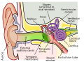
The ear or organ of hearing is divisible into three parts:
The external ear
The middle ear or tympanic cavity and
The internal ear or labyrinth
The Development of the Ear
The first rudiment of the internal ear appears shortly after that of the eye, in the form of a patch of thickened ectoderm, the auditory plate over the region of the hind-brain. The auditory plate becomes depressed and converted into the auditory pit (Fig. 898). The mouth of the pit is then closed, and thus a shut sac, the auditory vesicle is formed (Fig. 899); from it the epithelial lining of the membranous labyrinth is derived. The vesicle becomes pear-shaped, and the neck of the flask is obliterated (Fig. 900). From the vesicle certain diverticula are given off which form the various parts of the membranous labyrinth.
One from the middle part forms the ductus and saccus endolymphaticus, another from the anterior end gradually elongates, and, forming a tube coiled on itself, becomes the cochlear duct, the vestibular extremity of which is subsequently constricted to form the canalis reuniens. Three others appear as disk-like evaginations on the surface of the vesicle; the central parts of the walls of the disks coalesce and disappear, while the peripheral portions persist to form the semicircular ducts; of these the superior is the first and the lateral the last to be completed (Fig. 902). The central part of the vesicle represents the membranous vestibule, and is subdivided by a constriction into a smaller ventral part, the saccule, and a larger dorsal and posterior part, the utricle. This subdivision is effected by a fold which extends deeply into the proximal part of the ductus endolymphaticus, with the result that the utricle and saccule ultimately communicate with each other by means of a Y-shaped canal. The saccule opens into the cochlear duct, through the canalis reuniens, and the semicircular ducts communicate with the utricle.
The mesodermal tissue surrounding the various parts of the epithelial labyrinth is converted into a cartilaginous ear-capsule, and this is finally ossified to form the bony labyrinth. Between the cartilaginous capsule and the epithelial structures is a stratum of mesodermal tissue which is differentiated into three layers, viz., an outer, forming the periosteal lining of the bony labyrinth; an inner, in direct contact with the epithelial structures; and an intermediate, consisting of gelatinous tissue: by the absorption of this latter tissue the perilymphatic spaces are developed. The modiolus and osseous spiral lamina of the cochlea are not preformed in cartilage but are ossified directly from connective tissue.
The middle ear and auditory tube are developed from the first pharyngeal pouch. The entodermal lining of the dorsal end of this pouch is in contact with the ectoderm of the corresponding pharyngeal groove; by the extension of the mesoderm between these two layers the tympanic membrane is formed. During the sixth or seventh month the tympanic antrum appears as an upward and backward expansion of the tympanic cavity. With regard to the exact mode of development of the ossicles of the middle ear there is some difference of opinion.
The view generally held is that the malleus is developed from the proximal end of the mandibular (Meckel’s) cartilage (Fig. 43), the incus in the proximal end of the mandibular arch, and that the stapes is formed from the proximal end of the hyoid arch. The malleus, with the exception of its anterior process is ossified from a single center which appears near the neck of the bone; the anterior process is ossified separately in membrane and joins the main part of the bone about the sixth month of fetal life.
The incus is ossified from one center which appears in the upper part of its long crus and ultimately extends into its lenticular process. The stapes first appears as a ring (annulus stapedius) encircling a small vessel, the stapedial artery, which subsequently undergoes atrophy; it is ossified from a single center which appears in its base.
The external acoustic meatus is developed from the first branchial groove. The lower part of this groove extends inward as a funnel-shaped tube (primary meatus) from which the cartilaginous portion and a small part of the roof of the osseous portion of the meatus are developed. From the lower part of the funnel-shaped tube an epithelial lamina extends downward and inward along the inferior wall of the primitive tympanic cavity; by the splitting of this lamina the inner part of the meatus (secondary meatus) is produced, while the inner portion of the lamina forms the cutaneous stratum of the tympanic membrane.
The auricula or pinna is developed by the gradual differentiation of tubercles which appear around the margin of the first branchial groove. The rudiment of the acoustic nerve appears about the end of the third week as a group of ganglion cells closely applied to the cephalic edge of the auditory vesicle. Whether these cells are derived from the ectoderm adjoining the auditory vesicle, or have migrated from the wall of the neural tube, is as yet uncertain. The ganglion gradually splits into two parts, the vestibular ganglion and the spiral ganglion The peripheral branches of the vestibular ganglion pass in two divisions, the pars superior giving rami to the superior ampulla of the superior semicircular duct, to the lateral ampulla and to the utricle; and the pars inferior giving rami to the saccule and the posterior ampulla. The proximal fibers of the vestibular ganglion form the vestibular nerve; the proximal fibers of the spiral ganglion form the cochlear nerve.
Additional images
-
-
 Ear labyrinth
 -
Inner ear
-
Temporal bone


External links
 The dictionary definition of the organ of hearing at Wiktionary
| Anatomy of hearing and balance | ||||||||||||||||||||||||||||
|---|---|---|---|---|---|---|---|---|---|---|---|---|---|---|---|---|---|---|---|---|---|---|---|---|---|---|---|---|
|
| Human regional anatomy | ||||||||||
|---|---|---|---|---|---|---|---|---|---|---|
|
Gray's Anatomy
- Gray's Anatomy Contents
- Gray's Anatomy Subject Index
- About Classic Gray's Anatomy
- Glossary of anatomy terms
Anatomy atlases (external)
[1] - Anatomy Atlases
 | |
|---|---|
|
|
 |
| Human systems and organs | ||||||||||||||
|---|---|---|---|---|---|---|---|---|---|---|---|---|---|---|
|
