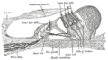
Stereocilia (inner ear)
Stereocilia in the inner ear are small, stiff, hair-like structures that are found on the top (apical surface) of hair cells in the organ of Corti and the vestibular system of the inner ear. They are not true cilia because they do not have the typical 9+2 structure of microtubules. Instead, they are filled with tightly packed actin filaments, which are anchored at the top by a dense body and at the base by a rootlet.
Structure and Function[edit]
Stereocilia are arranged in a staircase pattern of increasing height. They are interconnected by a series of tip links and horizontal top connectors. When sound vibrations or head movements cause the basilar membrane to move, this movement is transferred to the stereocilia, causing them to tilt and triggering the opening of mechanotransduction channels at the tips of the stereocilia. This allows potassium and calcium ions to flow into the hair cell, leading to depolarization and the release of neurotransmitters that stimulate the auditory nerve.
Clinical Significance[edit]
Damage to the stereocilia can lead to hearing loss and balance disorders. This can be caused by exposure to loud noise, certain medications (ototoxic drugs), and aging. In some cases, genetic mutations can also cause defects in the structure or function of the stereocilia, leading to congenital hearing loss.
See Also[edit]
References[edit]

This WikiMD article can only be edited by registered and verified editors. You can log in or register.
-
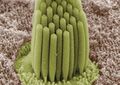 Stereocilia of frog inner ear
 -
 Stereocilia (inner ear)


")
Medical Disclaimer: WikiMD is for informational purposes only and is not a substitute for professional medical advice. Content may be inaccurate or outdated and should not be used for diagnosis or treatment. Always consult your healthcare provider for medical decisions. Verify information with trusted sources such as CDC.gov and NIH.gov. By using this site, you agree that WikiMD is not liable for any outcomes related to its content. See full disclaimer.
Credits:Most images are courtesy of Wikimedia commons, and templates, categories Wikipedia, licensed under CC BY SA or similar.
Translate page: - East Asian
中文,
日本,
한국어,
South Asian
हिन्दी,
தமிழ்,
తెలుగు,
Urdu,
ಕನ್ನಡ,
Southeast Asian
Indonesian,
Vietnamese,
Thai,
မြန်မာဘာသာ,
বাংলা
European
español,
Deutsch,
français,
Greek,
português do Brasil,
polski,
română,
русский,
Nederlands,
norsk,
svenska,
suomi,
Italian
Middle Eastern & African
عربى,
Turkish,
Persian,
Hebrew,
Afrikaans,
isiZulu,
Kiswahili,
Other
Bulgarian,
Hungarian,
Czech,
Swedish,
മലയാളം,
मराठी,
ਪੰਜਾਬੀ,
ગુજરાતી,
Portuguese,
Ukrainian
