Encephalocele: Difference between revisions
m 1 revision imported |
No edit summary |
||
| Line 1: | Line 1: | ||
{{Infobox medical condition (new) | {{Infobox medical condition (new) | ||
| name = Encephalocele | | name = Encephalocele | ||
| synonyms = Cranium bifidum | | synonyms = '''Cranium bifidum''' | ||
| image = Encephalocele-web.jpg | | image = Encephalocele-web.jpg | ||
| caption = Illustration of a child with encephalocele | | caption = Illustration of a child with encephalocele | ||
| pronounce = | | pronounce = | ||
| field = | | field = [[Neurosurgery]], [[Pediatrics]], [[Medical genetics]] | ||
| symptoms = | | symptoms = Visible sac-like protrusion on the head, [[hydrocephalus]], [[developmental delay]], [[seizures]], [[vision problems]] | ||
| complications = | | complications = [[Neurological deficits]], [[motor impairment]], [[intellectual disability]], [[infection]] | ||
| onset = | | onset = Present at [[birth]] | ||
| duration = | | duration = Lifelong (chronic condition) | ||
| types = | | types = [[Frontal encephalocele]], [[occipital encephalocele]], [[parietal encephalocele]], [[basal encephalocele]] | ||
| causes = | | causes = Failure of the [[neural tube]] to close completely during fetal development | ||
| risks = | | risks = [[Folate deficiency]], maternal exposure to [[teratogens]], family history of neural tube defects | ||
| diagnosis = | | diagnosis = [[Prenatal ultrasound]], [[MRI]] or [[CT scan]] after birth | ||
| differential = | | differential = [[Meningocele]], [[arachnoid cyst]], [[dermoid cyst]] | ||
| prevention = | | prevention = Adequate [[folic acid]] intake before and during pregnancy | ||
| treatment = | | treatment = Surgical repair, supportive therapy, [[shunt]] placement if hydrocephalus is present | ||
| medication = | | medication = Symptomatic management (e.g. [[antiepileptic drugs]] for seizures) | ||
| prognosis = | | prognosis = Depends on size, location, and brain involvement; can range from mild to severe disability | ||
| frequency = | | frequency = Rare; estimated incidence is 1 in 10,000 live births worldwide | ||
| deaths = | | deaths = Can be fatal if severe or untreated | ||
}} | }} | ||
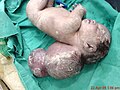
'''Encephalocele''', is a [[neural tube defect]] characterized by sac-like protrusions of the [[brain]] and the [[Biological membrane|membrane]]s that cover it through openings in the [[human skull|skull]]. These defects are caused by failure of the neural tube to close completely during fetal development. Encephaloceles cause a groove down the middle of the skull, or between the forehead and nose, or on the back side of the skull. The severity of encephalocele varies, depending on its location. | [[File:Ajcr-1-16.f1.jpg|Neonate with encephalocele|left|thumb]] | ||
'''Encephalocele''', is a [[neural tube defect]] characterized by sac-like protrusions of the [[brain]] and the [[Biological membrane|membrane]]s that cover it through openings in the [[human skull|skull]]. These defects are caused by failure of the neural tube to close completely during fetal development. Encephaloceles cause a groove down the middle of the skull, or between the forehead and nose, or on the back side of the skull. The severity of encephalocele varies, depending on its location. | |||
==Signs and symptoms== | ==Signs and symptoms== | ||
Encephaloceles are often accompanied by [[craniofacial abnormalities]] or other brain malformations. Symptoms may include neurologic problems, [[hydrocephalus]] (cerebrospinal fluid accumulated in the brain), [[spastic quadriplegia]] (paralysis of the limbs), [[microcephaly]] (an abnormally small head), [[ataxia]] (uncoordinated muscle movement), developmental delay, vision problems, mental and growth retardation, and seizures. | Encephaloceles are often accompanied by [[craniofacial abnormalities]] or other brain malformations. Symptoms may include neurologic problems, [[hydrocephalus]] (cerebrospinal fluid accumulated in the brain), [[spastic quadriplegia]] (paralysis of the limbs), [[microcephaly]] (an abnormally small head), [[ataxia]] (uncoordinated muscle movement), developmental delay, vision problems, mental and growth retardation, and seizures. | ||
==Causes== | ==Causes== | ||
| Line 50: | Line 43: | ||
==Treatment== | ==Treatment== | ||
Currently, the only effective treatment for encephaloceles is reparative surgery, generally performed during [[infancy]]. The extent to which it can be corrected depends on the location and size of the encephaloceles; however, large protrusions can be removed without causing major disability. Surgery repositions the bulging area back into the skull, removes the protrusions, and corrects the deformities, typically relieving pressure that can delay normal brain development. Occasionally, [[cerebral shunt|shunts]] are placed to drain excess cerebrospinal fluid from the brain. | Currently, the only effective treatment for encephaloceles is reparative surgery, generally performed during [[infancy]]. The extent to which it can be corrected depends on the location and size of the encephaloceles; however, large protrusions can be removed without causing major disability. Surgery repositions the bulging area back into the skull, removes the protrusions, and corrects the deformities, typically relieving pressure that can delay normal brain development. Occasionally, [[cerebral shunt|shunts]] are placed to drain excess cerebrospinal fluid from the brain. | ||
== Gallery == | |||
<gallery> | |||
File:Encephalocele of a newborn.JPG|A neonate with a large encephalocele. | |||
File:Encephalocele2.jpg|Encephalocele on the head of a two-year-old. | |||
File:Baby with encephalocele.jpg|Baby with encephalocele. | |||
File:Encephalocele picture.jpg|Encephalocele. | |||
</gallery> | |||
==See also== | ==See also== | ||
*[[Cephalic disorder]] | *[[Cephalic disorder]] | ||
*[[Knobloch syndrome]] | *[[Knobloch syndrome]] | ||
== External links == | == External links == | ||
| Line 101: | Line 71: | ||
}} | }} | ||
* {{NINDS|encephaloceles}} | * {{NINDS|encephaloceles}} | ||
{{Congenital malformations and deformations of nervous system}} | {{Congenital malformations and deformations of nervous system}} | ||
[[Category:Congenital disorders of nervous system]] | [[Category:Congenital disorders of nervous system]] | ||
[[Category:Disorders causing seizures]] | [[Category:Disorders causing seizures]] | ||
Latest revision as of 00:44, 27 March 2025
| Encephalocele | |
|---|---|

| |
| Synonyms | Cranium bifidum |
| Pronounce | |
| Field | Neurosurgery, Pediatrics, Medical genetics |
| Symptoms | Visible sac-like protrusion on the head, hydrocephalus, developmental delay, seizures, vision problems |
| Complications | Neurological deficits, motor impairment, intellectual disability, infection |
| Onset | Present at birth |
| Duration | Lifelong (chronic condition) |
| Types | Frontal encephalocele, occipital encephalocele, parietal encephalocele, basal encephalocele |
| Causes | Failure of the neural tube to close completely during fetal development |
| Risks | Folate deficiency, maternal exposure to teratogens, family history of neural tube defects |
| Diagnosis | Prenatal ultrasound, MRI or CT scan after birth |
| Differential diagnosis | Meningocele, arachnoid cyst, dermoid cyst |
| Prevention | Adequate folic acid intake before and during pregnancy |
| Treatment | Surgical repair, supportive therapy, shunt placement if hydrocephalus is present |
| Medication | Symptomatic management (e.g. antiepileptic drugs for seizures) |
| Prognosis | Depends on size, location, and brain involvement; can range from mild to severe disability |
| Frequency | Rare; estimated incidence is 1 in 10,000 live births worldwide |
| Deaths | Can be fatal if severe or untreated |

Encephalocele, is a neural tube defect characterized by sac-like protrusions of the brain and the membranes that cover it through openings in the skull. These defects are caused by failure of the neural tube to close completely during fetal development. Encephaloceles cause a groove down the middle of the skull, or between the forehead and nose, or on the back side of the skull. The severity of encephalocele varies, depending on its location.
Signs and symptoms[edit]
Encephaloceles are often accompanied by craniofacial abnormalities or other brain malformations. Symptoms may include neurologic problems, hydrocephalus (cerebrospinal fluid accumulated in the brain), spastic quadriplegia (paralysis of the limbs), microcephaly (an abnormally small head), ataxia (uncoordinated muscle movement), developmental delay, vision problems, mental and growth retardation, and seizures.
Causes[edit]
Although the exact cause is unknown, encephaloceles are caused by failure of the neural tube to close completely during fetal development. Research has indicated that teratogens (substances known to cause birth defects), trypan blue (a stain used to color dead tissues or cells blue), and arsenic may damage the developing fetus and cause encephaloceles. (August 2015)
Proper levels of folic acid have been shown to help prevent such defects when taken before pregnancy, and early in pregnancy.
Diagnosis[edit]
Usually encephaloceles are noticeable deformities and are diagnosed immediately after birth, but a small encephalocele in the nasal or forehead region can go undetected. Various physical and mental developmental delays can indicate the presence of encephaloceles.
Classifications[edit]
Encephaloceles of the face are generally classified as nasofrontal, nasoethmoidal, or naso-orbital, however, there can be some overlap in the type of encephalocele. They can also appear along any part of the cranial vault, as they result from abnormal closure of cranial bones; the most common location for encephaloceles is the occipital region. If the bulging portion contains only cerebrospinal fluid and the overlying membrane, it may be called a meningocele. If brain tissue is present, it may be referred to as a meningoencephalocele.<ref>If both brein tissue and ventricular cerebrospinal fluid are present, it may be called a meningohydroencephalocele.Encephalocele Imaging at eMedicine </ref> When the head size or occipitofrontal circumference is smaller than the herniating sac, then it is termed as giant encephalocele. <ref>9</ref>
Prevention[edit]
It is recommended that women who may become pregnant take 400 micrograms of folic acid daily. (December 2011)
Treatment[edit]
Currently, the only effective treatment for encephaloceles is reparative surgery, generally performed during infancy. The extent to which it can be corrected depends on the location and size of the encephaloceles; however, large protrusions can be removed without causing major disability. Surgery repositions the bulging area back into the skull, removes the protrusions, and corrects the deformities, typically relieving pressure that can delay normal brain development. Occasionally, shunts are placed to drain excess cerebrospinal fluid from the brain.
Gallery[edit]
-
 A neonate with a large encephalocele.
 -
 Encephalocele on the head of a two-year-old.
 -
 Baby with encephalocele.
 -
 Encephalocele.


See also[edit]
External links[edit]
| Congenital malformations and deformations of nervous system | ||||||||||
|---|---|---|---|---|---|---|---|---|---|---|
|
