YAP1: Difference between revisions
CSV import |
CSV import |
||
| Line 37: | Line 37: | ||
[[Category:Electromagnetic radiation]] | [[Category:Electromagnetic radiation]] | ||
[[Category:Radiology]] | [[Category:Radiology]] | ||
<gallery> | |||
File:Modular_Structure_of_YAP1_Isoforms.jpg|Modular Structure of YAP1 Isoforms | |||
File:YAP_and_TAZ_-_Biochemical_Regulation_Diagram.png|YAP and TAZ - Biochemical Regulation Diagram | |||
</gallery> | |||
Latest revision as of 02:15, 18 February 2025
X-ray[edit]
X-rays are a form of electromagnetic radiation, similar to visible light but with much higher energy and the ability to penetrate most substances to varying degrees. They are widely used in medicine, industry, and research.
History[edit]
X-rays were discovered in 1895 by the German physicist Wilhelm Conrad Röntgen. He was investigating the effects of cathode rays in a gas discharge tube when he noticed a fluorescent glow of crystals on a nearby table. Röntgen realized that a new type of ray, which he called "X" for unknown, was being emitted from the tube. His discovery revolutionized medical diagnostics and earned him the first Nobel Prize in Physics in 1901.
Properties[edit]
X-rays are part of the electromagnetic spectrum, with wavelengths ranging from about 0.01 to 10 nanometers. They have higher energy than ultraviolet light and can penetrate most materials, including human tissue. This property makes them invaluable for medical imaging.
Medical Applications[edit]
X-rays are primarily used in medicine for diagnostic imaging. The most common application is in radiography, where X-rays pass through the body and are captured on film or digital detectors to create images of the internal structures. This technique is essential for diagnosing fractures, infections, and tumors.
Computed Tomography (CT)[edit]
Computed Tomography (CT) is an advanced form of X-ray imaging that produces cross-sectional images of the body. It involves taking multiple X-ray measurements from different angles and using computer processing to create detailed images of the inside of the body.
Fluoroscopy[edit]
Fluoroscopy is a technique that uses X-rays to obtain real-time moving images of the interior of an object. It is often used during diagnostic and therapeutic procedures, such as catheter insertions and barium studies.
Safety[edit]
While X-rays are a powerful diagnostic tool, they do pose some risks due to their ionizing nature, which can damage living tissue. It is important to minimize exposure and use protective measures, such as lead aprons and thyroid shields, especially in repeated or high-dose procedures.
Industrial and Research Applications[edit]
Beyond medicine, X-rays are used in various industrial applications, such as inspecting welds and detecting structural flaws in materials. In research, X-ray crystallography is a technique used to determine the atomic and molecular structure of a crystal.
Also see[edit]
| Medical imaging | ||||||||||
|---|---|---|---|---|---|---|---|---|---|---|
* Category
|
| Electromagnetic radiation | ||||||||||
|---|---|---|---|---|---|---|---|---|---|---|
This electromagnetic radiation related article is a stub.
|
-
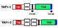 Modular Structure of YAP1 Isoforms
 -
 YAP and TAZ - Biochemical Regulation Diagram


