Computed tomography of the abdomen and pelvis
Computed tomography of the abdomen and pelvis is a medical imaging technique used to visualize the internal structures of the abdomen and pelvis. It uses computed tomography (CT) technology to generate detailed images of the organs, blood vessels, and tissues in these regions.
Overview[edit]
Computed tomography of the abdomen and pelvis is a non-invasive diagnostic procedure. It uses a combination of X-rays and computer technology to produce cross-sectional images (often called 'slices') of the body. These images provide more detailed information than conventional x-ray exams.
Procedure[edit]
During a CT scan of the abdomen and pelvis, the patient lies on a table that moves through a circular opening in the CT scanner. The scanner rotates around the patient and takes a series of images from different angles. These images are then processed by a computer to produce detailed cross-sectional images of the abdomen and pelvis.
Applications[edit]
Computed tomography of the abdomen and pelvis is used to diagnose and monitor a variety of health conditions, including cancer, inflammatory diseases, infections, and trauma. It can also be used to guide certain medical procedures, such as biopsies and drainages.
Risks[edit]
While CT scans are generally safe, they do expose the patient to a small amount of radiation. However, the benefits of an accurate diagnosis usually outweigh the risks. Other potential risks include allergic reactions to contrast material and kidney damage in patients with poor kidney function.
See also[edit]
This article related to medical imaging is a stub. You can help WikiMD by expanding the page. |
This radiology related article is a stub. You can help WikiMD by expanding it.
-
Computed tomography of the abdomen and pelvis
-
Ruptured abdominal aortic aneurysm
-
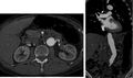
Wilms tumor CT scan
-
Non-contrast CT of multiple bilateral renal calculi
-
Normal contrast enhanced abdominal CT
-
 Abdominal CT angiography
 -
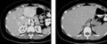
Arterial and portal venous phase CT of cholangiocarcinoma
-
 Late arterial and portal venous phase CT of focal nodular hyperplasia
 -
Corticomedullary phase CT in axial and coronal plane, and parenchymal phase, of renal cell carcinomaCorticomedullary phase CT in axial and coronal plane, and parenchymal phase, of renal cell carcinoma
-
Renal parenchymal phase CT of transitional cell carcinoma
-
Non-contrast, early arterial, and late arterial phase CT of pancreas with hypoenhancing mass


Medical Disclaimer: WikiMD is for informational purposes only and is not a substitute for professional medical advice. Content may be inaccurate or outdated and should not be used for diagnosis or treatment. Always consult your healthcare provider for medical decisions. Verify information with trusted sources such as CDC.gov and NIH.gov. By using this site, you agree that WikiMD is not liable for any outcomes related to its content. See full disclaimer.
Credits:Most images are courtesy of Wikimedia commons, and templates, categories Wikipedia, licensed under CC BY SA or similar.
Translate page: - East Asian
中文,
日本,
한국어,
South Asian
हिन्दी,
தமிழ்,
తెలుగు,
Urdu,
ಕನ್ನಡ,
Southeast Asian
Indonesian,
Vietnamese,
Thai,
မြန်မာဘာသာ,
বাংলা
European
español,
Deutsch,
français,
Greek,
português do Brasil,
polski,
română,
русский,
Nederlands,
norsk,
svenska,
suomi,
Italian
Middle Eastern & African
عربى,
Turkish,
Persian,
Hebrew,
Afrikaans,
isiZulu,
Kiswahili,
Other
Bulgarian,
Hungarian,
Czech,
Swedish,
മലയാളം,
मराठी,
ਪੰਜਾਬੀ,
ગુજરાતી,
Portuguese,
Ukrainian
