Jugular venous pressure: Difference between revisions
No edit summary |
CSV import |
||
| Line 35: | Line 35: | ||
[[Category:Internal medicine]] | [[Category:Internal medicine]] | ||
[[Category:Interventional cardiology]] | [[Category:Interventional cardiology]] | ||
== Jugular_venous_pressure == | |||
<gallery> | |||
File:Elevated_JVP.JPG|Elevated jugular venous pressure | |||
File:Gray558.png|Anatomy of the jugular vein | |||
File:JVP_waveform.png|Jugular venous pressure waveform | |||
</gallery> | |||
Latest revision as of 01:18, 18 February 2025
The Jugular Venous Pressure (JVP), occasionally referred to as the jugular venous pulse, represents an indirect evaluation of the pressure within the right atrium of the heart. Clinicians observe this pressure by inspecting the internal jugular vein, and it serves as a critical diagnostic tool for differentiating various types of heart and lung diseases.
Physiological Basis[edit]
The JVP arises from blood flowing back into the venous system, reflecting the pressure dynamics within the right atrium. It comprises of three upward (a, c, and v waves) and two downward deflections (x and y descents), each corresponding to specific physiological events within the cardiac cycle.
Clinical Assessment[edit]
JVP assessment is a standard part of the cardiovascular examination, providing information on fluid balance, right heart function, and central venous pressure. Clinicians usually measure it with the patient positioned at a 45-degree angle, identifying the highest point of oscillation of the internal jugular vein.
Pathophysiology and Clinical Significance[edit]
Alterations in JVP can indicate various pathophysiological conditions. Elevated JVP, for instance, may suggest right ventricular failure, tricuspid valve disease, or pulmonary hypertension. In contrast, a decreased JVP may indicate hypovolemia.
The specific wave patterns can also help identify heart conditions. For instance, a prominent 'a' wave might suggest tricuspid stenosis or right atrial myxoma, while large 'v' waves might indicate tricuspid regurgitation.
Limitations[edit]
While JVP is a valuable clinical indicator, it is not without limitations. Inter-observer variability, obesity, and patient positioning can affect its measurement accuracy.
References[edit]
<references/>
- 1. "Understanding jugular venous pressure." American Family Physician. [Link]
- 2. Colombo PC, Doran AC, Onat D, Wong F. (2020). "Clinical Assessment of the Jugular Venous Pressure." JACC Heart Fail. 8(12):1049-1051.
- 3. Cook DJ, Simel DL. (1996). "The Rational Clinical Examination. Does this patient have abnormal central venous pressure?" JAMA. 275(8):630-4.
- 4. Sarkisian AE. (2006). "Use of the physical examination to assess for volume status in heart failure patients." Curr Heart Fail Rep. 3(2):65-70.
 | |
|---|---|
|
|
 |
| Medical examination and history taking | ||||||||||||||||||
|---|---|---|---|---|---|---|---|---|---|---|---|---|---|---|---|---|---|---|
|
| Physiology of the cardiovascular system | ||||||||||||||
|---|---|---|---|---|---|---|---|---|---|---|---|---|---|---|
|
| Symptoms and signs relating to the cardiovascular system | ||||||||||
|---|---|---|---|---|---|---|---|---|---|---|
|
Jugular_venous_pressure[edit]
-
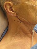 Elevated jugular venous pressure
 -
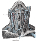 Anatomy of the jugular vein
 -
 Jugular venous pressure waveform


