Superior thoracic aperture


The superior thoracic aperture, also known anatomically as the thoracic inlet and clinically as the thoracic outlet, represents the upper opening of the thoracic cavity. This structure is crucial to understand, especially in the context of the clinical entity known as the Thoracic Outlet Syndrome. This syndrome pertains to the superior thoracic aperture and not its inferior counterpart.
Boundaries[edit]
The thoracic inlet is distinguished as an orifice encircled by a bony structure, facilitating the passage of several pivotal anatomical structures. The bounding features of the superior thoracic aperture are:
- Posteriorly: The first thoracic vertebra (T1).
- Laterally: The first pair of ribs, which form a C-shaped curve from the posterior to anterior.
- Anteriorly: The costal cartilage of the first rib coupled with the superior border of the manubrium.
Relations[edit]
The thoracic inlet's relational aspects are multifaceted:
- The clavicle joins with the manubrium, thereby demarcating the anterior perimeter of the thoracic inlet.
- Situated superior to the thoracic inlet is the neck's root, with the superior mediastinum positioned inferiorly.
- The brachial plexus has a superolateral relation to the thoracic inlet. This plexus arises between the anterior and middle scalene muscles, just above the first rib. It then transitions obliquely and inferiorly, passing beneath the clavicle towards the shoulder and subsequently, the arm. Thoracic outlet syndrome is often due to the brachial plexus becoming compressed in this region.
Contents[edit]
Structures that pass through the superior thoracic aperture include:
- trachea
- oesophagus
- thoracic duct
- apexes of the lungs
- nerves
- vessels
- arteries
- left and right common carotid arteries
- left and right subclavian arteries
- veins
- arteries
- lymph nodes and lymphatic vessels
Several structures traverse through the superior thoracic aperture, including:
Additionally, several minor yet vital nerves and vessels traverse through the aperture. In some scenarios, an enlarged thyroid gland might descend through the thoracic inlet, extending into the superior mediastinum.
The positioning of the oesophagus is adjacent to the body of the T1 vertebra, only separated by the prevertebral fascia. The trachea, residing anterior to the oesophagus, is centrally located and might make contact with the manubrium. The lungs' apices are positioned laterally to both the oesophagus and trachea, demarcated from the other structures by the mentioned vessels and nerves. They also project slightly above the inlet's level, for example, aligning with the first rib's horizontal plane.
See Also[edit]
Additional images[edit]
-
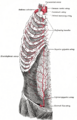 Vasculature entering at top. (Note: internal mammary is now known as internal thoracic artery.)

References[edit]
McMinn, RMH (Ed) (1994) Last's Anatomy: Regional and applied (9th Ed). London: Churchill Livingstone. ISBN 0-443-04662-X
This medical article is a stub. You can help WikiMD by expanding the page. |
| Bones of the torso | ||||||||||||||||
|---|---|---|---|---|---|---|---|---|---|---|---|---|---|---|---|---|
|
| Anatomy and morphology | ||||||||||
|---|---|---|---|---|---|---|---|---|---|---|
|
Medical Disclaimer: WikiMD is for informational purposes only and is not a substitute for professional medical advice. Content may be inaccurate or outdated and should not be used for diagnosis or treatment. Always consult your healthcare provider for medical decisions. Verify information with trusted sources such as CDC.gov and NIH.gov. By using this site, you agree that WikiMD is not liable for any outcomes related to its content. See full disclaimer.
Credits:Most images are courtesy of Wikimedia commons, and templates, categories Wikipedia, licensed under CC BY SA or similar.
Translate this page: - East Asian
中文,
日本,
한국어,
South Asian
हिन्दी,
தமிழ்,
తెలుగు,
Urdu,
ಕನ್ನಡ,
Southeast Asian
Indonesian,
Vietnamese,
Thai,
မြန်မာဘာသာ,
বাংলা
European
español,
Deutsch,
français,
Greek,
português do Brasil,
polski,
română,
русский,
Nederlands,
norsk,
svenska,
suomi,
Italian
Middle Eastern & African
عربى,
Turkish,
Persian,
Hebrew,
Afrikaans,
isiZulu,
Kiswahili,
Other
Bulgarian,
Hungarian,
Czech,
Swedish,
മലയാളം,
मराठी,
ਪੰਜਾਬੀ,
ગુજરાતી,
Portuguese,
Ukrainian
