The Vomer
Anatomy > Gray's Anatomy of the Human Body > II. [Osteology]] > 5b. 7. The Vomer
Henry Gray (1821–1865). Anatomy of the Human Body. 1918.
The Vomer[edit]
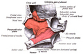
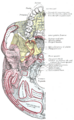
The vomer is situated in the median plane, but its anterior portion is frequently bent to one or other side. It is thin, somewhat quadrilateral in shape, and forms the hinder and lower part of the nasal septum (Fig. 173); it has two surfaces and four borders.
surfaces[edit]
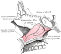
The surfaces (Fig. 174) are marked by small furrows for blood-vessels, and on each is the nasopalatine groove which runs obliquely downward and forward, and lodges the nasopalatine nerve and vessels.
Border[edit]
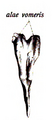
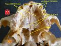
The superior border the thickest, presents a deep furrow, bounded on either side by a horizontal projecting ala of bone; the furrow receives the rostrum of the sphenoid, while the margins of the alae articulate with the vaginal processes of the medial pterygoid plates of the sphenoid behind, and with the sphenoidal processes of the palatine bones in front.
The inferior border articulates with the crest formed by the maxillae and palatine bones.
The anterior border is the longest and slopes downward and forward. Its upper half is fused with the perpendicular plate of the ethmoid; its lower half is grooved for the inferior margin of the septal cartilage of the nose.
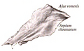
The posterior border is free, concave, and separates the choanae. It is thick and bifid above, thin below.

FIG. 173– Median wall of left nasal cavity showing vomer in situ (Picture From the Classic Gray's Anatomy)
Ossification[edit]
At an early period the septum of the nose consists of a plate of cartilage, the ethmovomerine cartilage The postero-superior part of this cartilage is ossified to form the perpendicular plate of the ethmoid; its antero-inferior portion persists as the septal cartilage, while the vomer is ossified in the membrane covering its postero-inferior part.
Two ossific centers, one on either side of the middle line, appear about the eighth week of fetal life in this part of the membrane, and hence the vomer consists primarily of two lamellae. About the third month these unite below, and thus a deep groove is formed in which the cartilage is lodged. As growth proceeds, the union of the lamellae extends upward and forward, and at the same time the intervening plate of cartilage undergoes absorption.
By the age of puberty the lamellae are almost completely united to form a median plate, but evidence of the bilaminar origin of the bone is seen in the everted alae of its upper border and the groove on its anterior margin.

FIG. 174– The vomer. (Picture From the Classic Gray's Anatomy)

FIG. 175– Vomer of infant. (Picture From the Classic Gray's Anatomy)
Articulations[edit]
The vomer articulates with six bones: two of the cranium, the sphenoid and ethmoid; and four of the face, the two maxillae and the two palatine bones; it also articulates with the septal cartilage of the nose.
Function[edit]
The vomeronasal organ, also called Jacobson's organ, is a chemoreceptor organ named for its closeness to the vomer and nasal bones, and is particularly developed in animals such as cats (who adopt a characteristic pose called the Flehmen reaction or flehming when making use of it), and is thought to have to do with the perception of certain pheromones.
Additional images[edit]
-
-
-
-
 Median wall of left nasal cavity showing vomer in situ.
 -
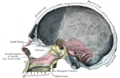
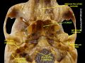
 Base of skull. Inferior surface.
 -
 Sagittal section of skull.
 -
-
 Vomer
Vomer -
 Vomer
Vomer


| The facial skeleton of the skull | ||||||||||||||||||||||||
|---|---|---|---|---|---|---|---|---|---|---|---|---|---|---|---|---|---|---|---|---|---|---|---|---|
|
Gray's Anatomy[edit]
- Gray's Anatomy Contents
- Gray's Anatomy Subject Index
- About Classic Gray's Anatomy
- Glossary of anatomy terms
Anatomy atlases (external)[edit]
[1] - Anatomy Atlases
| Human systems and organs | ||||||||||||||
|---|---|---|---|---|---|---|---|---|---|---|---|---|---|---|
|

Medical Disclaimer: WikiMD is for informational purposes only and is not a substitute for professional medical advice. Content may be inaccurate or outdated and should not be used for diagnosis or treatment. Always consult your healthcare provider for medical decisions. Verify information with trusted sources such as CDC.gov and NIH.gov. By using this site, you agree that WikiMD is not liable for any outcomes related to its content. See full disclaimer.
Credits:Most images are courtesy of Wikimedia commons, and templates, categories Wikipedia, licensed under CC BY SA or similar.
Translate page: - East Asian
中文,
日本,
한국어,
South Asian
हिन्दी,
தமிழ்,
తెలుగు,
Urdu,
ಕನ್ನಡ,
Southeast Asian
Indonesian,
Vietnamese,
Thai,
မြန်မာဘာသာ,
বাংলা
European
español,
Deutsch,
français,
Greek,
português do Brasil,
polski,
română,
русский,
Nederlands,
norsk,
svenska,
suomi,
Italian
Middle Eastern & African
عربى,
Turkish,
Persian,
Hebrew,
Afrikaans,
isiZulu,
Kiswahili,
Other
Bulgarian,
Hungarian,
Czech,
Swedish,
മലയാളം,
मराठी,
ਪੰਜਾਬੀ,
ગુજરાતી,
Portuguese,
Ukrainian
