File:Eye I.jpg
From WikiMD's medical encyclopedia

Size of this preview: 614 × 599 pixels. Other resolutions: 246 × 240 pixels | 492 × 480 pixels | 615 × 600 pixels | 787 × 768 pixels | 1,049 × 1,024 pixels | 2,098 × 2,048 pixels | 5,000 × 4,880 pixels.
Original file (5,000 × 4,880 pixels, file size: 17.81 MB, MIME type: image/jpeg)
Summary
| Description |
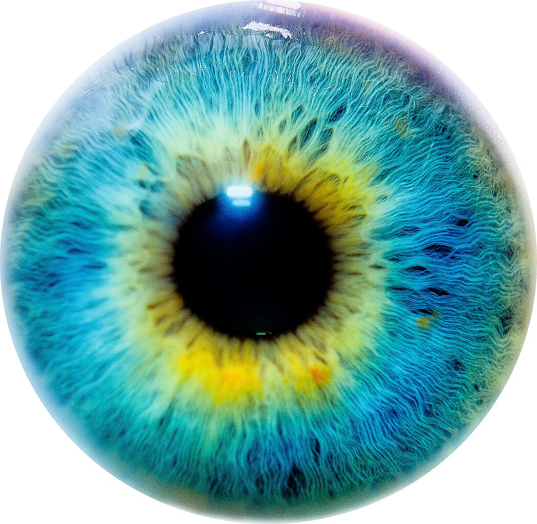
As seen by me. Facebook Fan Page | Twitter |
| Date | |
| Source | Flickr: Eye I |
| Author | Thomas Tolkien |
Licensing
This file is licensed under the Creative Commons Attribution 2.0 Generic license.
Attribution:
- You are free:
- to share – to copy, distribute and transmit the work
- to remix – to adapt the work
- Under the following conditions:
- attribution – You must give appropriate credit, provide a link to the license, and indicate if changes were made. You may do so in any reasonable manner, but not in any way that suggests the licensor endorses you or your use.
| This image, which was originally posted to Flickr, was uploaded to Commons using Flickr upload bot on 9 August 2011, 23:02 by Herkuleshippo. On that date, it was confirmed to be licensed under the terms of the license indicated. |
File history
Click on a date/time to view the file as it appeared at that time.
| Date/Time | Thumbnail | Dimensions | User | Comment | |
|---|---|---|---|---|---|
| current | 00:03, 21 November 2020 | | 5,000 × 4,880 (17.81 MB) | SteinsplitterBot | Bot: Image rotated by 180° |
File usage
The following 48 pages use this file:
- Accommodate
- Adaptation (eye)
- Amsler sign
- Anecortave acetate
- Argyll Robertson pupil
- Axenfeld Rieger syndrome
- Biological effects of high-energy visible light
- Blue field entoptic phenomenon
- Bruns nystagmus
- Buphthalmos
- Ciliary ganglion
- Clinical Ophthalmology (journal)
- Cogan syndrome
- Collier's sign
- Cornea plana 1
- Cyclic nucleotide-gated channel alpha 3
- Cyclospasm
- Dehydroretinal
- Diffuse lamellar keratitis
- Eales disease
- Entopic
- Fluocinolone acetonide
- Fundus (eye)
- Fundus photography
- Giant retinal ganglion cells
- Gonioscopy
- Hirschberg test
- Intraocular lens scaffold
- Intraocular pressure
- Iridocorneal endothelial syndrome
- Irvine–Gass syndrome
- Ispahani Islamia Eye Institute and Hospital
- Keratoendotheliitis fugax hereditaria
- Khodadoust line
- Lazy eye
- Lipaemia retinalis
- Listing's law
- NCX-466
- Pascal Photocoagulator
- Pellucid marginal degeneration
- Pholedrine
- Pupillometry
- Robert Salus
- Sclerotomy
- Snellen chart
- Suspensory ligament of eyeball
- Template:Ophthalmology
- Template:Ophthalmology-stub