Eye
The eye is the organ that allows us to perceive the sense of vision. The eye is a complex and sophisticated organ responsible for converting light into an electrochemical signal that can be processed by the brain, thereby allowing organisms to perceive visual information. As one of the most intricate organs in the human body, the eye provides the essential sense of vision which plays a fundamental role in myriad daily activities and interactions with the environment.

Anatomy of the Eye[edit]
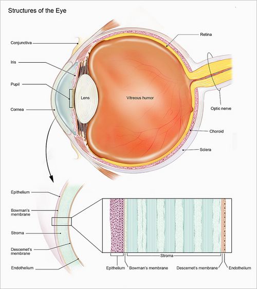
The eye is composed of multiple components, each serving a specific function in the process of vision.
- Sclera: The white outer layer of the eye that maintains its shape and protects its internal structures.
- Cornea: The clear, dome-shaped surface that covers the front of the eye and helps focus light.
- Iris: The colored part of the eye, which controls the size of the pupil and thus the amount of light entering the eye.
- Pupil: The black central opening in the iris that regulates the amount of light entering the eye.
- Lens: A flexible structure that adjusts its shape to focus light on the retina.
- Retina: A light-sensitive layer at the back of the eye where light is converted into electrical signals.
- Optic nerve: The nerve that transmits visual information from the retina to the brain.
Physiology of Vision[edit]
The process of vision begins when light rays enter the eye through the cornea, pass through the pupil, and then through the lens. The lens focuses this light onto the retina, where it produces an image. This image is detected by photoreceptor cells in the retina: the rods (sensitive to low light) and cones (sensitive to color). These cells convert the image into electrical signals which are then transmitted to the brain via the optic nerve. The brain then interprets these signals, allowing us to perceive the image as a visual scene.
Protection and Lubrication[edit]
The eye features several structures that safeguard and maintain it:
- Eyelids: Protect the eye from foreign objects and excessive light.
- Eyelashes: Trap dust and other small particles to keep them away from the eye.
- Tear glands: Produce tears that lubricate the eye and contain enzymes that break down bacteria.
- Conjunctiva: A thin layer covering the white of the eye and the inner side of the eyelids, providing additional protection and lubrication.
Common Disorders of the Eye[edit]
Several disorders can affect vision and the health of the eye. Some of the more common ones include:
- Myopia: Difficulty seeing distant objects.
- Hyperopia: Difficulty seeing close objects.
- Astigmatism: Distorted vision caused by an irregularly shaped cornea or lens.
- Cataract: Clouding of the eye's lens.
- Glaucoma: Damage to the optic nerve, usually due to increased eye pressure.
- Macular degeneration: A leading cause of vision loss affecting the central portion of the retina.
Research and Advances[edit]
Ophthalmologic research continues to unveil new insights into vision and ocular health. This includes work on gene therapies for inherited eye disorders, innovative surgical techniques, and the development of artificial retinas. In the realm of technological advancements, there's also a growing interest in devices and implants that can restore or enhance vision.
Cornea The cornea is the eye's outermost layer. It is the clear, dome-shaped surface that covers the front of the eye.
Common eye problems[edit]
Macular degeneration[edit]
- Dry form
The dry form of age-related macular degeneration (AMD) occurs when the light-sensitive cells in the macula slowly break down, gradually blurring central vision in the affected eye. Over time, as less of the macula functions, central vision is gradually lost in the affected eye.
- Wet form
The wet form of age-related macular degeneration occurs when abnormal blood vessels behind the retina start to grow under the macula. These new blood vessels tend to be very fragile and often leak blood and fluid. The blood and fluid raise the macula from its normal place at the back of the eye. Damage to the macula occurs rapidly.
Floaters[edit]
Floaters are tiny spots, specks, flecks and "cobwebs" that drift aimlessly around in your field of vision.
Lens[edit]
The lens is the clear part of the eye that helps to focus light, or an image, on the retina.
Macula[edit]
The macula is the part of the eye that allows you to see fine detail. The macula is located in the center of the retina.
Optic Nerve[edit]
The optic nerve sends visual information from the retina to the brain.
Retina[edit]
The retina is the light-sensitive tissue at the back of the eye. The retina instantly converts light, or an image, into electrical impulses. The retina then sends these impulses, or nerve signals, to the brain.
See also[edit]

Topics in Ophthalmology[edit]
- Macular degeneration (AMD)
- Amblyopia
- Anophthalmia and * Microphthalmia
- Astigmatism
- Blepharitis
- Cataract
- Color blindness
- Cornea and Corneal disease
- Diabetic retinopathy
- Dry eye
- Floaters
- Glaucoma
- Hyperopia
- Intracranial hypertension
- Low vision
- Macular edema
- Myopia
- Pink Eye or Conjunctivitis
- Presbyopia
- Refractive errors
- Retinal detachment
- Retinitis pigmentosa
- Retinoblastoma
- Retinopathy of prematurity
- Uveitis
- Vitreous detachment
Ad. Transform your life with W8MD's Budget GLP-1 injections from $49.99


W8MD offers a medical weight loss program to lose weight in Philadelphia. Our physician-supervised medical weight loss provides:
- Weight loss injections in NYC (generic and brand names):
- Zepbound / Mounjaro, Wegovy / Ozempic, Saxenda
- Most insurances accepted or discounted self-pay rates. We will obtain insurance prior authorizations if needed.
- Generic GLP1 weight loss injections from $49.99 for the starting dose of Semaglutide and $65.00 for Tirzepatide.
- Also offer prescription weight loss medications including Phentermine, Qsymia, Diethylpropion, Contrave etc.
NYC weight loss doctor appointmentsNYC weight loss doctor appointments
Start your NYC weight loss journey today at our NYC medical weight loss and Philadelphia medical weight loss clinics.
- Call 718-946-5500 to lose weight in NYC or for medical weight loss in Philadelphia 215-676-2334.
- Tags:NYC medical weight loss, Philadelphia lose weight Zepbound NYC, Budget GLP1 weight loss injections, Wegovy Philadelphia, Wegovy NYC, Philadelphia medical weight loss, Brookly weight loss and Wegovy NYC
Error creating thumbnail: ![]() Error creating thumbnail:
Error creating thumbnail:
![]()
|
WikiMD's Wellness Encyclopedia |
| Let Food Be Thy Medicine Medicine Thy Food - Hippocrates |
Medical Disclaimer: WikiMD is not a substitute for professional medical advice. The information on WikiMD is provided as an information resource only, may be incorrect, outdated or misleading, and is not to be used or relied on for any diagnostic or treatment purposes. Please consult your health care provider before making any healthcare decisions or for guidance about a specific medical condition. WikiMD expressly disclaims responsibility, and shall have no liability, for any damages, loss, injury, or liability whatsoever suffered as a result of your reliance on the information contained in this site. By visiting this site you agree to the foregoing terms and conditions, which may from time to time be changed or supplemented by WikiMD. If you do not agree to the foregoing terms and conditions, you should not enter or use this site. See full disclaimer.
Credits:Most images are courtesy of Wikimedia commons, and templates, categories Wikipedia, licensed under CC BY SA or similar.
Translate this page: - East Asian
中文,
日本,
한국어,
South Asian
हिन्दी,
தமிழ்,
తెలుగు,
Urdu,
ಕನ್ನಡ,
Southeast Asian
Indonesian,
Vietnamese,
Thai,
မြန်မာဘာသာ,
বাংলা
European
español,
Deutsch,
français,
Greek,
português do Brasil,
polski,
română,
русский,
Nederlands,
norsk,
svenska,
suomi,
Italian
Middle Eastern & African
عربى,
Turkish,
Persian,
Hebrew,
Afrikaans,
isiZulu,
Kiswahili,
Other
Bulgarian,
Hungarian,
Czech,
Swedish,
മലയാളം,
मराठी,
ਪੰਜਾਬੀ,
ગુજરાતી,
Portuguese,
Ukrainian


