Autoimmune pancreatitis
| Autoimmune pancreatitis | |
|---|---|
| [[File:|250px|alt=|]] | |
| Synonyms | AIP |
| Pronounce | |
| Field | |
| Symptoms | |
| Complications | |
| Onset | |
| Duration | |
| Types | |
| Causes | |
| Risks | |
| Diagnosis | |
| Differential diagnosis | |
| Prevention | |
| Treatment | |
| Medication | |
| Prognosis | |
| Frequency | |
| Deaths | |
Autoimmune pancreatitis is an increasingly recognized type of chronic pancreatitis that can be difficult to distinguish from pancreatic carcinoma but which responds to treatment with corticosteroids, particularly prednisone.<ref>,
 The autoimmune diseases,
 4th edition,
 Academic Press,
 2006,


 ISBN 978-0-12-595961-2,</ref> There are two categories of AIP: Type 1 and Type 2, each with distinct clinical profiles.
Type 1 AIP is now regarded as a manifestation of IgG4-related disease,<ref name=pmid22736240>,
 Recommendations for the nomenclature of IgG4-related disease and its individual organ system manifestations,
 Arthritis & Rheumatism,
 2012,
 Vol. 64(Issue: 10),
 pp. 3061–7,
 DOI: 10.1002/art.34593,
 PMID: 22736240,
 PMC: 5963880,</ref> and those affected have tended to be older and to have a high relapse rate. Type 1 is associated with pancreatitis, Sjogren syndrome, Primary sclerosing cholangitis and Inflammatory bowel disease.  Patients with Type 2 AIP do not experience relapse, tend to be younger and not associated with systemic disease. AIP occurring in association with an autoimmune disorder has been referred to as "secondary" or "syndromic" AIP. AIP does not affect long-term survival.<ref>,
 Differences in Clinical Profile and Relapse Rate of Type 1 Versus Type 2 Autoimmune Pancreatitis,
 Gastroenterology,
 2010,
 Vol. 139(Issue: 1),
 pp. 140–8; quiz e12–3,
 DOI: 10.1053/j.gastro.2010.03.054,
 PMID: 20353791,</ref>
Signs and symptoms
AIP is relatively uncommon<ref>,
 Diagnosis of Autoimmune Pancreatitis: The Mayo Clinic Experience,
 Clinical Gastroenterology and Hepatology,
 2006,
 Vol. 4(Issue: 8),
 pp. 1010–6; quiz 934,
 DOI: 10.1016/j.cgh.2006.05.017,
 PMID: 16843735,</ref> and is characterized by the following features:
- Scleral Icterus (yellow eyes), jaundice (yellow skin) which is usually painless, usually without acute attacks of pancreatitis.
- Relatively mild symptoms, such as minimal weight loss or nausea.
- Increased serum levels of gamma globulins, immunoglobulin G (IgG) or IgG4.
- The presence of serum autoantibodies such as anti-nuclear antibody (ANA), anti-lactoferrin antibody, anti-carbonic anhydrase II antibody, and rheumatoid factor (RF).
- Contrast-enhanced CT demonstrates a diffusely enlarged (sausage-shaped) pancreas.
- Diffuse irregular narrowing of the main pancreatic duct, and stenosis of the intrapancreatic bile duct on endoscopic retrograde cholangiopancreatography (ERCP).
- Rare pancreatic calcification or cyst formation.
- Marked responsiveness to treatment with corticosteroids.
Two-thirds of patients present with either obstructive painless jaundice or a "mass" in the head of the pancreas mimicking carcinoma. It is mandatory to rule out carcinoma prior to making a diagnosis of AIP. (October 2016)
Histopathology
Histopathologic examination of the pancreas reveals a characteristic lymphoplasmacytic infiltrate of CD4- or CD8-positive lymphocytes and IgG4-positive plasma cells, and exhibits interstitial fibrosis and acinar cell atrophy in later stages. At the initial stages, typically, there is a cuff of lymphoplasma cells surrounding the ducts but also more diffuse infiltration in the lobular parenchyma. However, localization and the degree of duct wall infiltration are variable. Whereas histopathologic examination remains the primary method for differentiation of AIP from acute and chronic pancreatitis, lymphoma, and cancer. By Fine Needle Aspiration (FNA) the diagnosis can be made if adequate tissue is obtained. In such cases, lymphoplasmacytic infiltration of the lobules are the key finding. Rarely, granulomatous reaction could be observed. It has been proposed that a cytologic smear primarily composed of acini rich in chronic inflammatory cells (lymphocytes, plasma cells), with rare ductal epithelial cells lacking atypia, favors the diagnosis of AIP. The sensitivity and the specificity of these criteria for differentiating AIP from neoplasia are unknown. In cases of systemic manifestation of AIP, the pathologic features would be similar in other organs. (October 2016)
Diagnosis
Criteria
Most recently the fourteenth Congress of the International Association of Pancreatology developed the International Consensus Diagnostic Criteria (ICDC) for AIP. The ICDC emphasizes five cardinal features of AIP which includes the imaging appearance of pancreatic parenchyma and the pancreatic duct, serum IgG4 level, other organ involvement with IgG4-related disease, pancreatic histology and response to steroid therapy.<ref name=pmid24758653>,
 Recent Advances in the Diagnosis and Management of Autoimmune Pancreatitis,
 American Journal of Roentgenology,
 2014,
 Vol. 202(Issue: 5),
 pp. 1007–21,
 DOI: 10.2214/AJR.13.11247,
 PMID: 24758653,</ref>
In 2002, the Japanese Pancreas Society proposed the following diagnostic criteria for autoimmune pancreatitis: (October 2016)
- I. Pancreatic imaging studies show diffuse narrowing of the main pancreatic duct with irregular wall (more than 1/3 of length of the entire pancreas).
- II. Laboratory data demonstrate abnormally elevated levels of serum gamma globulin and/or IgG, or the presence of autoantibodies.
- III. Histopathologic examination of the pancreas shows fibrotic changes with lymphocyte and plasma cell infiltrate.
For diagnosis, criterion I (pancreatic imaging) must be present with criterion II (laboratory data) and/or III (histopathologic findings).<ref>,
 How to diagnose autoimmune pancreatitis by the revised Japanese clinical criteria,
 Journal of Gastroenterology,
 2007,
 Vol. 42 Suppl 18,
 pp. 32–8,
 DOI: 10.1007/s00535-007-2049-5,
 PMID: 17520221,</ref>
Mayo Clinic has come up with five diagnostic criteria called HISORt criteria which stands for histology, imaging, serology, other organ involvement, and response to steroid therapy.<ref>,
 Review of the diagnosis, classification and management of autoimmune pancreatitis,
 World Journal of Gastrointestinal Pathophysiology,
 2014,
 Vol. 5(Issue: 2),
 pp. 71–81,
 DOI: 10.4291/wjgp.v5.i2.71,
 PMID: 24891978,
 PMC: 4025075,</ref>
Radiologic features
Computed tomography (CT) findings in AIP include a diffusely enlarged hypodense pancreas or a focal mass that may be mistaken for a pancreatic malignancy.<ref name=pmid24758653/> A low-density, capsule-like rim on CT (possibly corresponding to an inflammatory process involving peripancreatic tissues) is thought to be an additional characteristic feature (thus the mnemonic: sausage-shaped). Magnetic resonance imaging (MRI) reveals a diffusely decreased signal intensity and delayed enhancement on dynamic scanning. The characteristic ERCP finding is segmental or diffuse irregular narrowing of the main pancreatic duct, usually accompanied by an extrinsic-appearing stricture of the distal bile duct. Changes in the extrapancreatic bile duct similar to those of primary sclerosing cholangitis (PSC) have been reported. (October 2016)
The role of endoscopic ultrasound (EUS) and EUS-guided fine-needle aspiration (EUS-FNA) in the diagnosis of AIP is not well described, and EUS findings have been described in only a small number of patients. In one study, EUS revealed a diffusely swollen and hypoechoic pancreas in 8 of the 14 (57%) patients, and a solitary, focal, irregular mass was observed in 6 (46%) patients. Whereas EUS-FNA is sensitive and specific for the diagnosis of pancreatic malignancy, its role in the diagnosis of AIP remains unclear.
(October 2016)
Treatment
AIP often completely resolves with steroid treatment. The failure to differentiate AIP from malignancy may lead to unnecessary pancreatic resection, and the characteristic lymphoplasmacytic infiltrate of AIP has been found in up to 23% of patients undergoing pancreatic resection for suspected malignancy who are ultimately found to have benign disease. In this subset of patients, a trial of steroid therapy may have prevented a Whipple procedure or complete pancreatectomy for a benign disease which responds well to medical therapy.<ref>,
 Autoimmune Chronic Pancreatitis,
 Journal of the Chinese Medical Association,
 2008,
 Vol. 71(Issue: 1),
 pp. 14–22,
 DOI: 10.1016/S1726-4901(08)70067-4,
 PMID: 18218555,</ref> "This benign disease resembles pancreatic carcinoma both clinically and radiographically. The diagnosis of autoimmune pancreatitis is challenging to make. However, accurate and timely diagnosis may preempt the misdiagnosis of cancer and decrease the number of unnecessary pancreatic resections."<ref name="ReferenceA">,
 Autoimmune pancreatitis: A mimic of pancreatic cancer,
 Cleveland Clinic Journal of Medicine,
 2009,
 Vol. 76(Issue: 10),
 pp. 607–15,
 DOI: 10.3949/ccjm.76a.09039,
 PMID: 19797461,</ref> Autoimmune pancreatitis responds dramatically to corticosteroid treatment.<ref name="ReferenceA"/>
If relapse occurs after corticosteroid treatment or corticosteroid treatment is not tolerated, immunomodulators may be used. Immunomodulators such as azathioprine, and 6-mercaptopurine have been shown to extend remission of autoimmune pancreatitis after corticosteroid treatment. If corticosteroid and immunomodulator treatments are not sufficient, rituximab may also be used. Rituximab has been shown to induce and maintain remission.<ref>,
 Immunomodulators and Rituximab in the Management of Autoimmune Pancreatitis,
 Pancreapedia,
 2013,


 DOI: 10.3998/panc.2013.20,</ref>
Controversies in nomenclature
As the number of published cases of AIP has increased, efforts have been focused on defining AIP as a distinct clinical and pathologic entity and toward developing some generally agreed upon diagnostic criteria and nomenclature. Terms frequently encountered are autoimmune or autoimmune-related pancreatitis, lymphoplasmacytic sclerosing pancreatitis, idiopathic tumefactive chronic pancreatitis, idiopathic pancreatitis with focal irregular narrowing of the main pancreatic duct, and non-alcoholic duct destructive chronic pancreatitis. There are also a large number of case reports employing descriptive terminology such as pancreatitis associated with Sjögren’s syndrome, primary sclerosing cholangitis, or inflammatory bowel disease. Some of the earliest cases were reported as pancreatic pseudotumor or pseudolymphoma. (October 2016)
References
External links

-
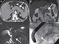 Diffuse autoimmune pancreatitis

Ad. Transform your life with W8MD's
GLP-1 weight loss injections special from $29.99


W8MD Medical Weight Loss, Sleep and Medspa offers physician-supervised medical weight loss programs: NYC medical weight loss Philadelphia medical weight loss
Affordable GLP-1 Weight Loss ShotsAffordable GLP-1 Weight Loss Shots
Budget GLP-1 injections NYC (insurance & self-pay options) Popular treatments:
- Semaglutide starting from $29.99/week
- Tirzepatide starting from $45.00/week
✔ Most insurances accepted for visits ✔ Prior authorization support when eligible
Start your physician weight loss NYC journey today:
📍 NYC: Brooklyn weight loss center 📍 Philadelphia: Philadelphia weight loss center
📞 Call: 718-946-5500 (NYC) | 215-676-2334 (Philadelphia)
Tags: Affordable GLP1 weight loss NYC, Wegovy NYC, Zepbound NYC, Philadelphia medical weight loss
![]()
![]()
![]()
![]()
|
WikiMD Medical Encyclopedia |
Medical Disclaimer: WikiMD is not a substitute for professional medical advice. The information on WikiMD is provided as an information resource only, may be incorrect, outdated or misleading, and is not to be used or relied on for any diagnostic or treatment purposes. Please consult your health care provider before making any healthcare decisions or for guidance about a specific medical condition. WikiMD expressly disclaims responsibility, and shall have no liability, for any damages, loss, injury, or liability whatsoever suffered as a result of your reliance on the information contained in this site. By visiting this site you agree to the foregoing terms and conditions, which may from time to time be changed or supplemented by WikiMD. If you do not agree to the foregoing terms and conditions, you should not enter or use this site. See full disclaimer.
Credits:Most images are courtesy of Wikimedia commons, and templates, categories Wikipedia, licensed under CC BY SA or similar.
Translate this page: - East Asian
中文,
日本,
한국어,
South Asian
हिन्दी,
தமிழ்,
తెలుగు,
Urdu,
ಕನ್ನಡ,
Southeast Asian
Indonesian,
Vietnamese,
Thai,
မြန်မာဘာသာ,
বাংলা
European
español,
Deutsch,
français,
Greek,
português do Brasil,
polski,
română,
русский,
Nederlands,
norsk,
svenska,
suomi,
Italian
Middle Eastern & African
عربى,
Turkish,
Persian,
Hebrew,
Afrikaans,
isiZulu,
Kiswahili,
Other
Bulgarian,
Hungarian,
Czech,
Swedish,
മലയാളം,
मराठी,
ਪੰਜਾਬੀ,
ગુજરાતી,
Portuguese,
Ukrainian
