Umbilical folds: Difference between revisions
CSV import |
CSV import |
||
| Line 42: | Line 42: | ||
[[Category:Anatomy of the abdomen]] | [[Category:Anatomy of the abdomen]] | ||
<gallery> | |||
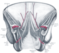
File:Gray1036.png|Umbilical folds | |||
File:Inguinal_fossae.PNG|Inguinal fossae | |||
</gallery> | |||
Latest revision as of 01:57, 17 February 2025
Anatomical structures in the human abdomen
| Anatomy and morphology | ||||||||||
|---|---|---|---|---|---|---|---|---|---|---|
|
Umbilical folds[edit]
The umbilical folds are anatomical structures located on the internal surface of the anterior abdominal wall. They are peritoneal folds that cover the underlying structures and are important landmarks in the abdomen.
Types[edit]
There are five umbilical folds:
- The median umbilical fold is a single midline structure that covers the urachus, a remnant of the fetal connection between the bladder and the umbilicus.
- The medial umbilical folds are paired structures that cover the obliterated umbilical arteries.
- The lateral umbilical folds are also paired and cover the inferior epigastric vessels.
Function[edit]
The umbilical folds serve as important landmarks during laparoscopic surgery and other medical procedures involving the abdominal cavity. They help in identifying the location of the inguinal fossae and other structures.
Inguinal fossae[edit]
The inguinal fossae are depressions on the internal surface of the anterior abdominal wall, located lateral to the umbilical folds. They are clinically significant as they are potential sites for inguinal hernias.
Types[edit]
There are three types of inguinal fossae:
- The supravesical fossa is located between the median and medial umbilical folds.
- The medial inguinal fossa is situated between the medial and lateral umbilical folds.
- The lateral inguinal fossa is found lateral to the lateral umbilical fold and is the site of the deep inguinal ring.
Clinical significance[edit]
The umbilical folds and inguinal fossae are important in the diagnosis and treatment of hernias. The lateral inguinal fossa, in particular, is the site where indirect inguinal hernias occur, while direct inguinal hernias occur through the medial inguinal fossa.
Images[edit]


Related pages[edit]
References[edit]
- Moore, Keith L.; Dalley, Arthur F.; Agur, Anne M. R. (2013). Clinically Oriented Anatomy. 7th edition. Lippincott Williams & Wilkins.
- Standring, Susan (2015). Gray's Anatomy: The Anatomical Basis of Clinical Practice. 41st edition. Elsevier.
-
 Umbilical folds
 -
 Inguinal fossae
