Ardipithecus ramidus: Difference between revisions
CSV import |
CSV import Tags: mobile edit mobile web edit |
||
| Line 29: | Line 29: | ||
[[Category:Cardiovascular system]] | [[Category:Cardiovascular system]] | ||
[[Category:Embryology]] | [[Category:Embryology]] | ||
==Ardipithecus ramidus== | |||
<gallery> | |||
File:Ardipithecus_ramidus.jpg|Ardipithecus ramidus | |||
File:Map_of_the_fossil_sites_of_the_earliest_hominids_(35.8-3.3M_BP).svg|Map of the fossil sites of the earliest hominids | |||
File:Ardipithecus_Gesamt1.jpg|Ardipithecus ramidus | |||
File:Modell_eines_Schädels_des_Pan_troglodytes_(Schimpanse,_weiblich).jpg|Model of a female chimpanzee skull | |||
File:Nut_cracking_Ardipithecus.png|Nut cracking Ardipithecus | |||
</gallery> | |||
Latest revision as of 04:59, 18 February 2025
Structure in the developing heart
| Anatomy and morphology | ||||||||||
|---|---|---|---|---|---|---|---|---|---|---|
|
The aorticopulmonary septum is a critical structure in the developing heart of the embryo. It plays a vital role in the separation of the aorta and the pulmonary artery, which are the major arteries that carry blood away from the heart. This septum is essential for the proper division of the outflow tract of the heart into the systemic and pulmonary circulations.
Development[edit]
The aorticopulmonary septum develops from the neural crest cells and the truncus arteriosus during embryogenesis. The process begins with the migration of neural crest cells into the truncus arteriosus, which is the common arterial trunk that initially serves both the systemic and pulmonary circulations.
As development progresses, these cells contribute to the formation of the spiral septum, which divides the truncus arteriosus into the aorta and the pulmonary trunk. This division is crucial for establishing the separate pathways for oxygenated and deoxygenated blood, which is a hallmark of the mammalian circulatory system.
Function[edit]
The primary function of the aorticopulmonary septum is to ensure that the aorta and the pulmonary artery are properly aligned with their respective ventricles. The aorta must connect to the left ventricle, which pumps oxygenated blood to the body, while the pulmonary artery must connect to the right ventricle, which pumps deoxygenated blood to the lungs.
Clinical significance[edit]
Defects in the formation of the aorticopulmonary septum can lead to congenital heart defects such as persistent truncus arteriosus, where the septum fails to form properly, resulting in a single arterial trunk. Other related conditions include transposition of the great arteries and tetralogy of Fallot, which involve improper alignment or separation of the aorta and pulmonary artery.
Related pages[edit]
Gallery[edit]
-
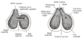 Diagram showing the development of the heart in the human embryo.
 -
 Illustration of the aorticopulmonary septum in the developing heart.

Ardipithecus ramidus[edit]
-
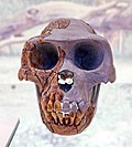 Ardipithecus ramidus
 -
 Map of the fossil sites of the earliest hominids
 -
 Ardipithecus ramidus
 -
 Model of a female chimpanzee skull
 -
 Nut cracking Ardipithecus

.svg)

.jpg)

