The Nasal Bones
Anatomy > Gray's Anatomy of the Human Body > II. [Osteology]] > 5b. The Facial Bones. 1. The Nasal Bones
Henry Gray (1821–1865). Anatomy of the Human Body. 1918.

The Nasal Bones[edit]
(Ossa Faciei) & (Ossa Nasalia)
The nasal bones are two small oblong bones, varying in size and form in different individuals; they are placed side by side at the middle and upper part of the face, and form, by their junction, “the bridge” of the nose (Fig. 190). Each has two surfaces and four borders.

Surfaces[edit]
The outer surface (Fig. 155) is concavoconvex from above downward, convex from side to side; it is covered by the Procerus and Compressor naris, and perforated about its center by a foramen, for the transmission of a small vein.
The inner surface (Fig. 156) is concave from side to side, and is traversed from above downward, by a groove for the passage of a branch of the nasociliary nerve.
Borders[edit]
The superior border is narrow, thick, and serrated for articulation with the nasal notch of the frontal bone. The inferior border is thin, and gives attachment to the lateral cartilage of the nose; near its middle is a notch which marks the end of the groove just referred to.
The lateral border is serrated, bevelled at the expense of the inner surface above, and of the outer below, to articulate with the frontal process of the maxilla.
The medial border thicker above than below, articulates with its fellow of the opposite side, and is prolonged behind into a vertical crest, which forms part of the nasal septum: this crest articulates, from above downward, with the spine of the frontal, the perpendicular plate of the ethmoid, and the septal cartilage of the nose.

Ossification[edit]
Each bone is ossified from one center, which appears at the beginning of the third month of fetal life in the membrane overlying the front part of the cartilaginous nasal capsule.
Articulations[edit]
The nasal articulates with four bones: two of the cranium, the frontal and ethmoid, and two of the face, the opposite nasal and the maxilla.
Additional images[edit]
-
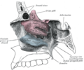 Lateral wall of nasal cavity, showing ethmoid bone in position.

External links[edit]
- Anatomy figure: 22:02-07 at Human Anatomy Online, SUNY Downstate Medical Center—"Anterior view of skull."
- Anatomy photo:29:st-0206 at the SUNY Downstate Medical Center—"Orbits and Eye: Bones"
- Anatomy figure: 33:01-03 at Human Anatomy Online, SUNY Downstate Medical Center—"The bones of the lateral nasal wall."
- Anatomy diagram: 34256.000-1. Elsevier.
| The facial skeleton of the skull | ||||||||||||||||||||||||
|---|---|---|---|---|---|---|---|---|---|---|---|---|---|---|---|---|---|---|---|---|---|---|---|---|
|
Gray's Anatomy[edit]
- Gray's Anatomy Contents
- Gray's Anatomy Subject Index
- About Classic Gray's Anatomy
- Glossary of anatomy terms
Anatomy atlases (external)[edit]
[1] - Anatomy Atlases
| Human systems and organs | ||||||||||||||
|---|---|---|---|---|---|---|---|---|---|---|---|---|---|---|
|

Medical Disclaimer: WikiMD is for informational purposes only and is not a substitute for professional medical advice. Content may be inaccurate or outdated and should not be used for diagnosis or treatment. Always consult your healthcare provider for medical decisions. Verify information with trusted sources such as CDC.gov and NIH.gov. By using this site, you agree that WikiMD is not liable for any outcomes related to its content. See full disclaimer.
Credits:Most images are courtesy of Wikimedia commons, and templates, categories Wikipedia, licensed under CC BY SA or similar.
Translate page: - East Asian
中文,
日本,
한국어,
South Asian
हिन्दी,
தமிழ்,
తెలుగు,
Urdu,
ಕನ್ನಡ,
Southeast Asian
Indonesian,
Vietnamese,
Thai,
မြန်မာဘာသာ,
বাংলা
European
español,
Deutsch,
français,
Greek,
português do Brasil,
polski,
română,
русский,
Nederlands,
norsk,
svenska,
suomi,
Italian
Middle Eastern & African
عربى,
Turkish,
Persian,
Hebrew,
Afrikaans,
isiZulu,
Kiswahili,
Other
Bulgarian,
Hungarian,
Czech,
Swedish,
മലയാളം,
मराठी,
ਪੰਜਾਬੀ,
ગુજરાતી,
Portuguese,
Ukrainian
