Phycomycosis

Editor-In-Chief: Prab R Tumpati, MD
Obesity, Sleep & Internal medicine
Founder, WikiMD Wellnesspedia &
W8MD's weight loss doctor NYC
Philadelphia GLP-1 weight loss and GLP-1 clinic NYC
| Phycomycosis | |
|---|---|

| |
| Synonyms | Zygomycosis, Mucormycosis |
| Pronounce | N/A |
| Specialty | N/A |
| Symptoms | Fever, skin lesions, sinusitis, pulmonary infection, gastrointestinal infection |
| Complications | Tissue necrosis, organ failure, sepsis |
| Onset | Rapid |
| Duration | Variable |
| Types | N/A |
| Causes | Fungal infection by Zygomycetes |
| Risks | Diabetes mellitus, immunocompromised state, iron overload |
| Diagnosis | Histopathology, culture, PCR |
| Differential diagnosis | Aspergillosis, Candidiasis, Bacterial infection |
| Prevention | Avoidance of exposure, control of underlying conditions |
| Treatment | Antifungal medication, surgical debridement |
| Medication | Amphotericin B, Posaconazole, Isavuconazole |
| Prognosis | Variable, often poor without treatment |
| Frequency | Rare |
| Deaths | N/A |
Phycomycosis is a type of fungal infection that affects both humans and animals. It is caused by various species of fungi in the order Mucorales. The disease is characterized by the formation of large, non-healing ulcers, often with a necrotic center and raised, indurated margins.
Etiology[edit]
Phycomycosis is caused by various species of fungi in the order Mucorales. These fungi are ubiquitous in the environment and are commonly found in soil, decaying organic matter, and the gastrointestinal tract of animals. Infection usually occurs through inhalation of fungal spores or direct inoculation into the skin or mucous membranes.
Clinical Presentation[edit]
The clinical presentation of phycomycosis can vary depending on the site of infection. Cutaneous phycomycosis presents as large, non-healing ulcers, often with a necrotic center and raised, indurated margins. Pulmonary phycomycosis can present with cough, fever, and hemoptysis. Gastrointestinal phycomycosis can present with abdominal pain, nausea, vomiting, and diarrhea.
Diagnosis[edit]
Diagnosis of phycomycosis is based on clinical presentation, histopathological examination, and culture of the fungus. Histopathological examination typically shows broad, non-septate hyphae with right-angle branching. Culture of the fungus can be performed on Sabouraud's agar.
Treatment[edit]
Treatment of phycomycosis involves surgical debridement of the infected tissue and antifungal therapy. The antifungal drug of choice is amphotericin B. In severe cases, combination therapy with other antifungal drugs may be required.
Prognosis[edit]
The prognosis of phycomycosis is generally poor, especially in immunocompromised patients. Early diagnosis and aggressive treatment can improve the prognosis.
Gallery[edit]
-
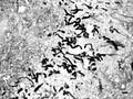 Hyphae of Pythium insidiosum
 -
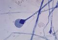 Mature sporangium of a Mucor species fungus


See Also[edit]


Medical Disclaimer: WikiMD is for informational purposes only and is not a substitute for professional medical advice. Content may be inaccurate or outdated and should not be used for diagnosis or treatment. Always consult your healthcare provider for medical decisions. Verify information with trusted sources such as CDC.gov and NIH.gov. By using this site, you agree that WikiMD is not liable for any outcomes related to its content. See full disclaimer.
Credits:Most images are courtesy of Wikimedia commons, and templates, categories Wikipedia, licensed under CC BY SA or similar.
Translate page: - East Asian
中文,
日本,
한국어,
South Asian
हिन्दी,
தமிழ்,
తెలుగు,
Urdu,
ಕನ್ನಡ,
Southeast Asian
Indonesian,
Vietnamese,
Thai,
မြန်မာဘာသာ,
বাংলা
European
español,
Deutsch,
français,
Greek,
português do Brasil,
polski,
română,
русский,
Nederlands,
norsk,
svenska,
suomi,
Italian
Middle Eastern & African
عربى,
Turkish,
Persian,
Hebrew,
Afrikaans,
isiZulu,
Kiswahili,
Other
Bulgarian,
Hungarian,
Czech,
Swedish,
മലയാളം,
मराठी,
ਪੰਜਾਬੀ,
ગુજરાતી,
Portuguese,
Ukrainian
