Gel electrophoresis of nucleic acids
Gel electrophoresis of nucleic acids is a fundamental technique used in molecular biology for the separation of DNA, RNA, or nucleic acid fragments based on their size. This method involves applying an electric field to move the nucleic acids through a gel matrix, allowing for the analysis of genetic material in research and clinical settings.
Principle[edit]
The principle behind gel electrophoresis of nucleic acids relies on the negative charge of the phosphate backbone of DNA and RNA molecules. When an electric field is applied, these negatively charged molecules migrate towards the positive electrode. The gel, typically made of agarose or polyacrylamide, acts as a molecular sieve, separating the nucleic acids by size: smaller fragments move faster and travel further than larger ones.
Types of Gel Electrophoresis[edit]
There are two main types of gel electrophoresis used for nucleic acids:
- Agarose Gel Electrophoresis: Primarily used for the separation of larger DNA fragments, ranging from 100 base pairs to over 20,000 base pairs. Agarose gels are easy to prepare and handle, making them suitable for most routine applications.
- Polyacrylamide Gel Electrophoresis (PAGE): Offers higher resolution than agarose gels and is typically used for the separation of smaller DNA or RNA fragments, up to 500 base pairs. PAGE is also the method of choice for the analysis of RNA secondary structure and DNA-protein interactions.
Procedure[edit]
The general procedure for gel electrophoresis of nucleic acids includes the following steps:
1. Preparation of the gel: Agarose or polyacrylamide gel is prepared and poured into a casting tray to solidify. 2. Loading of samples: Nucleic acid samples, along with a DNA ladder (a mixture of DNA fragments of known size), are loaded into the wells of the gel. 3. Electrophoresis: The gel is placed in an electrophoresis tank, covered with a buffer solution, and an electric current is applied. The nucleic acids migrate through the gel matrix. 4. Visualization: After electrophoresis, the gel is stained with a DNA-binding dye (e.g., ethidium bromide) to visualize the separated nucleic acid fragments under UV light.
Applications[edit]
Gel electrophoresis of nucleic acids is widely used in molecular biology and genetics for various applications, including:
- Analysis of PCR products
- Genotyping
- DNA sequencing preparation
- RNA analysis and RNA integrity assessment
- Verification of genetic engineering and cloning experiments
Safety Considerations[edit]
When performing gel electrophoresis of nucleic acids, it is important to follow safety guidelines due to the use of potentially hazardous materials, such as ethidium bromide, and the involvement of electrical equipment.
See Also[edit]
This molecular biology article is a stub. You can help WikiMD by expanding the page. |
-
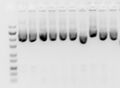
 DNA agarose gel electrophoresis
 -
 Electropherogram trace
 -
 Plasmid miniprep
 -
 Gel electrophoresis of nucleic acids
 -
 Agarose gel
 -
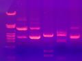
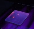 Agarose gel under UV light
 -
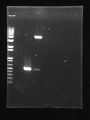 Agarose gel photo


Medical Disclaimer: WikiMD is for informational purposes only and is not a substitute for professional medical advice. Content may be inaccurate or outdated and should not be used for diagnosis or treatment. Always consult your healthcare provider for medical decisions. Verify information with trusted sources such as CDC.gov and NIH.gov. By using this site, you agree that WikiMD is not liable for any outcomes related to its content. See full disclaimer.
Credits:Most images are courtesy of Wikimedia commons, and templates, categories Wikipedia, licensed under CC BY SA or similar.
Translate page: - East Asian
中文,
日本,
한국어,
South Asian
हिन्दी,
தமிழ்,
తెలుగు,
Urdu,
ಕನ್ನಡ,
Southeast Asian
Indonesian,
Vietnamese,
Thai,
မြန်မာဘာသာ,
বাংলা
European
español,
Deutsch,
français,
Greek,
português do Brasil,
polski,
română,
русский,
Nederlands,
norsk,
svenska,
suomi,
Italian
Middle Eastern & African
عربى,
Turkish,
Persian,
Hebrew,
Afrikaans,
isiZulu,
Kiswahili,
Other
Bulgarian,
Hungarian,
Czech,
Swedish,
മലയാളം,
मराठी,
ਪੰਜਾਬੀ,
ગુજરાતી,
Portuguese,
Ukrainian
