The Optic Nerve
Anatomy > Gray's Anatomy of the Human Body > IX. Neurology > 5b. The Optic Nerve
Henry Gray (1821–1865). Anatomy of the Human Body. 1918.
The Optic Nerve
(N. Opticus; Second Nerve)
The optic nerve (Fig. 773), or nerve of sight consists mainly of fibers derived from the ganglionic cells of the retina. These axons terminate in arborizations around the cells in the lateral geniculate body, pulvinar, and superior colliculus which constitute the lower or primary visual centers.
From the cells of the lateral geniculate body and the pulvinar fibers pass to the cortical visual center, situated in the cuneus and in the neighborhood of the calcarine fissure. A few fibers of the optic nerve, of small caliber, pass from the primary centers to the retina and are supposed to govern chemical changes in the retina and also the movements of some of its elements (pigment cells and cones).
There are also a few fine fibers, afferent fibers, extending from the retina to the brain, that are supposed to be concerned in pupillary reflexes.
FIG. 773– The left optic nerve and the optic tracts. (Picture From the Classic Gray's Anatomy)
The optic nerve is peculiar in that its fibers and ganglion cells are probably third in the series of neurons from the receptors to the brain. Consequently the optic nerve corresponds rather to a tract of fibers within the brain than to the other cranial nerves. Its fibers pass backward and medialward through the orbit and optic foramen to the optic commissure where they partially decussate. The mixed fibers from the two nerves are continued in the optic tracts, the
primary visual centers of the brain
The orbital portion of the optic nerve is from 20 mm. to 30 mm. in length and has a slightly sinuous course to allow for movements of the eyeball. It is invested by an outer sheath of dura mater and an inner sheath from the arachnoid which are attached to the sclera around the area where the nerve fibers pierce the choroid and sclera of the bulb.
A little behind the bulb of the eye the central artery of the retina with its accompanying vein perforates the optic nerve, and runs within it to the retina. As the nerve enters the optic foramen its dural sheath becomes continuous with that lining the orbit and the optic foramen.
In the optic foramen the ophthalmic artery lies below and to its outer side. The intercranial portion of the optic nerve is about 10 mm. in length. The Optic Chiasma (chiasma opticum), somewhat quadrilateral in form, rests upon the tuberculum sellae and on the anterior part of the diaphragma sellae.
It is in relation, above with the lamina terminalis; behind with the tuber cinereum; on either side with the anterior perforated substance. Within the chiasma, the optic nerves undergo a partial decussation.
The fibers forming the medial part of each tract and posterior part of the chiasma have no connection with the optic nerves. They simply cross in the chiasma, and connect the medial geniculate bodies of the two sides; they form the commissure of Gudden The remaining and principal part of the chiasma consists of two sets of fibers, crossed and uncrossed.
The crossed fibers which are the more numerous, occupy the central part of the chiasma, and pass from the optic nerve of one side to the optic tract of the other, decussating in the chiasma with similar fibers of the opposite optic nerve.
The uncrossed fibers occupy the lateral part of the chiasma, and pass from the nerve of one side into the tract of the same side.
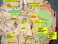
FIG. 774– Scheme showing central connections of the optic nerves and optic tracts. (Picture From the Classic Gray's Anatomy)
The crossed fibers of the optic nerve tend to occupy the medial side of the nerve and the uncrossed fibers the lateral side. In the optic tract, however, the fibers are much more intermingled.
Optic Tract
The Optic Tract (Fig. 774), passes backward and outward from the optic chiasma over the tuber cinereum and anterior perforated space to the cerebral peduncle and winds obliquely across its under surface.
Its fibers terminate in the lateral geniculate body, the pulvinar and the superior colliculus. It is adherent to the tuber cinereum and the cerebral peduncle as it passes over them. In the region of the lateral geniculate body it splits into two bands.
The medial and smaller one is a part of the commissure of Gudden and ends in the medial geniculate body. From its mode of development, and from its structure, the optic nerve must be regarded as a prolongation of the brain substance, rather than as an ordinary cerebrospinal nerve.
As it passes from the brain it receives sheaths from the three cerebral membranes, a perineural sheath from the pia mater, an intermediate sheath from the arachnoid, and an outer sheath from the dura mater, which is also connected with the periosteum as it passes through the optic foramen.
These sheaths are separated from each other by cavities which communicate with the subdural and subarachnoid cavities respectively. The innermost or perineural sheath sends a process around the arteria centralis retinae into the interior of the nerve, and enters intimately into its structure.
Note 130 A specimen of congenital absence of the optic chiasma is to be found in the Museum of the Westminister Hospital. See also Henle, Nervenlehre p. 393, ed. 2.
Function
The optic nerve transmits all visual information including brightness perception, color perception and contrast (visual acuity). It also conducts the visual impulses that are responsible for two important neurological reflexes: the light reflex and the accommodation reflex. The light reflex refers to the constriction of both pupils that occurs when light is shone into either eye. The accommodation reflex refers to the swelling of the lens of eye that occurs when one looks at a near object (for example, when reading the lens adjusts to near vision.
The eye's blind spot is a result of the absence of photoreceptors in the area of the retina where the optic nerve leaves the eye.<ref name="Vilensky" />
Additional images
-
 MRI scan of human eye showing optic nerve.
 -
 The ophthalmic artery derived from internal carotid artery and its branches. (optic nerve is yellow)
 -
 Superficial dissection of brain-stem. Lateral view.
 -
 Dissection of brain-stem. Lateral view.
 -
 Scheme showing central connections of the optic nerves and optic tracts.
 -
 Nerves of the orbit. Seen from above.
 -
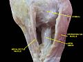
 Nerves of the orbit, and the ciliary ganglion. Side view.
 -
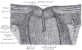 The terminal portion of the optic nerve and its entrance into the eyeball, in horizontal section.
 -
 Structures of the eye labeled
 -
 This image shows another labeled view of the structures of the eye
 -
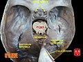
 Optic nerve.Deep dissection.Inferior view.
 -
 Optic nerve.Deep dissection.Inferior view.
 -
 Optic nerve
 -
 Optic nerve
 -
 Human brain dura mater (reflections)
 -
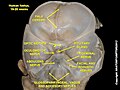
 Optic nerve
 -
 Optic nerve
 -
 Optic nerve
 -
 Cerebrum.Inferior view.Deep dissection
 -
Cerebral peduncle, optic chasm, cerebral aqueduct. Inferior view. Deep dissection.


_description.JPG)


External links
- The optic nerve on MRI
- Template:BrainMaps
- IFOND
- online case history – Optic nerve analysis with both scanning laser polarimetry with variable corneal compensation (GDx VCC) and confocal scanning laser ophthalmoscopy (HRT II - Heidelberg Retina Tomograph). Also includes actual fundus photos.
- Animations of extraocular cranial nerve and muscle function and damage (University of Liverpool)
- lesson3 at The Anatomy Lesson by Wesley Norman (Georgetown University)
(orbit4
)
- cranialnerves at The Anatomy Lesson by Wesley Norman (Georgetown University)
(II
)
| The cranial nerves | ||||||||||
|---|---|---|---|---|---|---|---|---|---|---|
|
| Optical illusions (list) | ||||||
|---|---|---|---|---|---|---|
|
Gray's Anatomy
- Gray's Anatomy Contents
- Gray's Anatomy Subject Index
- About Classic Gray's Anatomy
- Glossary of anatomy terms
Anatomy atlases (external)
[1] - Anatomy Atlases
 | |
|---|---|
|
|
 |
| Human systems and organs | ||||||||||||||
|---|---|---|---|---|---|---|---|---|---|---|---|---|---|---|
|
<references />
Ad. Transform your life with W8MD's Budget GLP-1 injections from $29.99


W8MD offers medical weight loss programs including NYC medical weight loss and Philadelphia medical weight loss offering:
- Affordable GLP1 shots (generic and brand names) such as
- Wegovy NYC (Semaglutide)
- Zepbound NYC /
- Learn more: Budget GLP1 weight loss injections NYC & Philadelphia GLP1 weight loss shots
- Most insurances accepted
- Lowest cost GLP1 weight loss NYC such as Semaglutide starting from $29.99/week and $45.00/week (Tirzepatide) with insurance.
- Prescription weight loss NYC including:
NYC weight loss doctor appointmentsNYC weight loss doctor appointments
Start your physician weight loss journey today at our:
- NYC medical weight loss
- Philadelphia medical weight loss
- Call 718-946-5500 for NYC or 215-676-2334 for Philadelphia
- Tags:
Budget glp1 weight loss NYC,
Zepbound NYC,
Philadelphia medical weight loss,
Wegovy NYC,
Budget Zepbound Philadelphia,
[** https://w8md.org/?page_id=62216 Affordable glp1 shots Philadelphia]
![]()
![]()
![]()
![]()
Advertise on WikiMD
|
WikiMD's Wellness Encyclopedia |
| Let Food Be Thy Medicine Medicine Thy Food - Hippocrates |
Medical Disclaimer: WikiMD is not a substitute for professional medical advice. The information on WikiMD is provided as an information resource only, may be incorrect, outdated or misleading, and is not to be used or relied on for any diagnostic or treatment purposes. Please consult your health care provider before making any healthcare decisions or for guidance about a specific medical condition. WikiMD expressly disclaims responsibility, and shall have no liability, for any damages, loss, injury, or liability whatsoever suffered as a result of your reliance on the information contained in this site. By visiting this site you agree to the foregoing terms and conditions, which may from time to time be changed or supplemented by WikiMD. If you do not agree to the foregoing terms and conditions, you should not enter or use this site. See full disclaimer.
Credits:Most images are courtesy of Wikimedia commons, and templates, categories Wikipedia, licensed under CC BY SA or similar.
Translate this page: - East Asian
中文,
日本,
한국어,
South Asian
हिन्दी,
தமிழ்,
తెలుగు,
Urdu,
ಕನ್ನಡ,
Southeast Asian
Indonesian,
Vietnamese,
Thai,
မြန်မာဘာသာ,
বাংলা
European
español,
Deutsch,
français,
Greek,
português do Brasil,
polski,
română,
русский,
Nederlands,
norsk,
svenska,
suomi,
Italian
Middle Eastern & African
عربى,
Turkish,
Persian,
Hebrew,
Afrikaans,
isiZulu,
Kiswahili,
Other
Bulgarian,
Hungarian,
Czech,
Swedish,
മലയാളം,
मराठी,
ਪੰਜਾਬੀ,
ગુજરાતી,
Portuguese,
Ukrainian
