The Parathyroid Glands
Anatomy > Gray's Anatomy of the Human Body > XI. Splanchnology > 4b. The Parathyroid Glands
Henry Gray (1821–1865). Anatomy of the Human Body. 1918.
The Parathyroid Glands
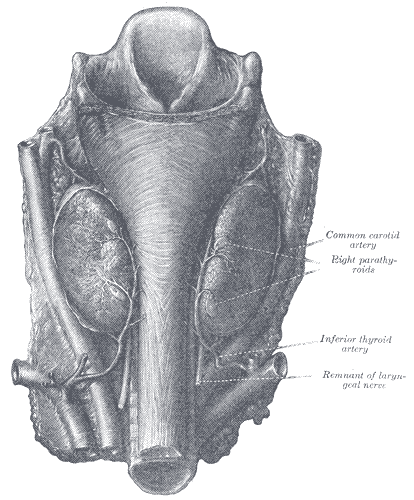
The parathyroid glands (Fig. 1177) are small brownish-red bodies, situated as a rule between the posterior borders of the lateral lobes of the thyroid gland and its capsule. They differ from it in structure, being composed of masses of cells arranged in a more or less columnar fashion with numerous intervening capillaries. They measure on an average about 6 mm. in length, and from 3 to 4 mm. in breadth, and usually present the appearance of flattened oval disks.
They are divided, according to their situation, into superior and inferior
The superior, usually two in number, are the more constant in position, and are situated, one on either side, at the level of the lower border of the cricoid cartilage, behind the junction of the pharynx and esophagus.
The inferior, also usually two in number, may be applied to the lower edge of the lateral lobes, or placed at some little distance below the thyroid gland, or found in relation to one of the inferior thyroid veins.
FIG. 1177– Parathyroid glands. (Halsted and Evans.) (Picture From the Classic Gray's Anatomy)
In man, they number four as a rule; fewer than four were found in less than 1 per cent. of over a thousand persons (Pepere 182), but more than four in over 33 per cent. of 122 bodies examined by Civalleri. In addition, numerous minute islands of parathyroid tissue may be found scattered in the connective tissue and fat of the neck around the parathyroid glands proper, and quite distinct from them.
Development
The parathyroid bodies are developed as outgrowths from the third and fourth branchial pouches (Figs. 1175). A pair of diverticula arise from the fifth branchial pouch and form what are termed the ultimo-branchial bodies (Fig. 1175): these fuse with the thyroid gland, but probably contribute no true thyroid tissue.
Structure
Microscopically the parathyroids consist of intercommunicating columns of cells supported by connective tissue containing a rich supply of blood capillaries. Most of the cells are clear, but some, larger in size, contain oxyphil granules. Vesicles containing colloid have been described as occurring in the parathyroid, but the observation has not been confirmed. No doubt the parathyroid glands produce an internal secretion essential to the well-being of the human economy; but it is still a matter of dispute what symptoms of disease are produced by their removal and suppression of their secretion. Pepere believes that they show signs of exceptional activity during pregnancy, and that parathyroid insufficiency is a main factor in the production of tetany in infants and adults, of eclampsia, and of certain sorts of fits. It is probable that the tetany following parathyroidectomy is due to the accumulation of ammonium carbonate and Kendall has suggested that the function of the parathyroid is to convert ammonium carbonate into urea.
Note 181 Consult an article “Concerning the Parathyroid Glands,” by D. A. Welsh, Journal of Anatomy and Physiology, vol. xxxii. Note 182 Consult Le Ghiandole paratiroidee, by A. Pepere, Turin, 1906.
Function
The major function of the parathyroid glands is to maintain the body's calcium and phosphate levels within a very narrow range, so that the nervous and muscular systems can function properly. The parathyroid glands do this by secreting parathyroid hormone (PTH).<ref name=WHEATERS2006p336>Barbara,
 Wheater's functional histology: a text and colour atlas,
 5th edition,
 Edinburgh:Churchill Livingstone/Elsevier,
 2006,


 ISBN 978-0-443-06850-8,</ref>
Parathyroid hormone (also known as parathormone) is a small protein that takes part in the control of calcium and phosphate homeostasis, as well as bone physiology. Parathyroid hormone has effects antagonistic to those of calcitonin.<ref name=GUYTONHALL2005>Arthur C. Guyton, John E.,
 Textbook of medical physiology,
 11th edition,
 Philadelphia:W.B. Saunders,
 2005,


 ISBN 978-0-7216-0240-0,


 Pages: 985–8,</ref>
- Calcium. PTH increases blood calcium levels by directly stimulating osteoblasts and thereby indirectly stimulating osteoclasts (through RANK/RANKL mechanism) to break down bone and release calcium. PTH increases gastrointestinal calcium absorption by activating vitamin D, and promotes calcium conservation (reabsorption) by the kidneys.<ref name=GUYTONHALL2005 />
- Phosphate. PTH is the major regulator of serum phosphate concentrations via actions on the kidney. It is an inhibitor of proximal tubular reabsorption of phosphorus. Through activation of vitamin D the absorption (intestinal) of Phosphate is increased.<ref name=GUYTONHALL2005 />
Additional images
-
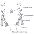 Scheme showing development of branchial epithelial bodies. I, II, III, IV. Branchial pouches.

External links
| Anatomy of the endocrine system | ||||||||
|---|---|---|---|---|---|---|---|---|
|
Gray's Anatomy
- Gray's Anatomy Contents
- Gray's Anatomy Subject Index
- About Classic Gray's Anatomy
- Glossary of anatomy terms
Anatomy atlases (external)
[1] - Anatomy Atlases
 | |
|---|---|
|
|
 |
| Human systems and organs | ||||||||||||||
|---|---|---|---|---|---|---|---|---|---|---|---|---|---|---|
|
Ad. Transform your health with W8MD Weight Loss, Sleep & MedSpa

Tired of being overweight?
Get started with evidence based, physician-supervised
affordable GLP-1 weight loss injections
Now available in New York City and Philadelphia:
- Semaglutide starting from $59.99/week and up
- Tirzepatide starting from $69.99/week and up (dose dependent)
✔ Evidence-based medical weight loss ✔ Insurance-friendly visits available ✔ Same-week appointments, evenings & weekends
Learn more:
Start your transformation today with W8MD weight loss centers.
|
WikiMD Medical Encyclopedia |
Medical Disclaimer: WikiMD is for informational purposes only and is not a substitute for professional medical advice. Content may be inaccurate or outdated and should not be used for diagnosis or treatment. Always consult your healthcare provider for medical decisions. Verify information with trusted sources such as CDC.gov and NIH.gov. By using this site, you agree that WikiMD is not liable for any outcomes related to its content. See full disclaimer.
Credits:Most images are courtesy of Wikimedia commons, and templates, categories Wikipedia, licensed under CC BY SA or similar.
Translate this page: - East Asian
中文,
日本,
한국어,
South Asian
हिन्दी,
தமிழ்,
తెలుగు,
Urdu,
ಕನ್ನಡ,
Southeast Asian
Indonesian,
Vietnamese,
Thai,
မြန်မာဘာသာ,
বাংলা
European
español,
Deutsch,
français,
Greek,
português do Brasil,
polski,
română,
русский,
Nederlands,
norsk,
svenska,
suomi,
Italian
Middle Eastern & African
عربى,
Turkish,
Persian,
Hebrew,
Afrikaans,
isiZulu,
Kiswahili,
Other
Bulgarian,
Hungarian,
Czech,
Swedish,
മലയാളം,
मराठी,
ਪੰਜਾਬੀ,
ગુજરાતી,
Portuguese,
Ukrainian
