The Popliteal Fossa: Difference between revisions
CSV import |
No edit summary |
||
| Line 33: | Line 33: | ||
[[Category:Lower limb anatomy]] | [[Category:Lower limb anatomy]] | ||
[[Category:Knee]] | [[Category:Knee]] | ||
{{grays}} | {{grays}} | ||
Latest revision as of 17:20, 4 February 2025
Anatomy > Gray's Anatomy of the Human Body > VI. The Arteries > 6b. The Popliteal Fossa
Henry Gray (1821–1865). Anatomy of the Human Body. 1918.
The Popliteal Fossa[edit]

Boundaries[edit]
The popliteal fossa (Fig. 551) or space is a lozenge-shaped space, at the back of the knee-joint. Laterally it is bounded by the Biceps femoris above, and by the Plantaris and the lateral head of the Gastrocnemius below; medially it is limited by the Semitendinous and Semimembranosus above, and by the medial head of the Gastrocnemius below. The floor is formed by the popliteal surface of the femur, the oblique popliteal ligament of the knee-joint, the upper end of the tibia, and the fascia covering the Popliteus; the fossa is covered in by the fascia lata.
Contents[edit]
The popliteal fossa contains the popliteal vessels, the tibial and the common peroneal nerves, the termination of the small saphenous vein, the lower part of the posterior femoral cutaneous nerve, the articular branch from the obturator nerve, a few small lymph glands, and a considerable quantity of fat.
The tibial nerve descends through the middle of the fossa, lying under the deep fascia and crossing the vessels posteriorly from the lateral to the medial side.
The common peroneal nerve descends on the lateral side of the upper part of the fossa, close to the tendon of the Biceps femoris. On the floor of the fossa are the popliteal vessels, the vein being superficial to the artery and united to it by dense areolar tissue; the vein is a thick-walled vessel, and lies at first lateral to the artery, and then crosses it posteriorly to gain its medial side below; sometimes it is double, the artery lying between the two veins, which are usually connected by short transverse branches.
The articular branch from the obturator nerve descends upon the artery to the knee-joint. The popliteal lymph glands, six or seven in number, are imbedded in the fat; one lies beneath the popliteal fascia near the termination of the external saphenous vein, another between the popliteal artery and the back of the knee-joint, while the others are placed at the sides of the popliteal vessel. Arising from the artery, and passing off from it at right angles, are its genicular branches.
Additional images[edit]
-
 Muscles of the gluteal and posterior femoral regions.
 -
 Small saphenous vein and its tributaries.
 -
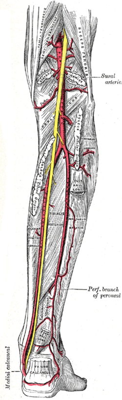 The popliteal, posterior tibial, and peroneal arteries.
 -
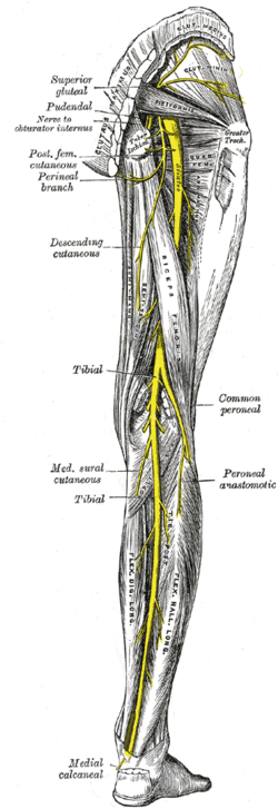 Nerves of the right lower extremity Posterior view.
 -
 Muscles of thigh. Lateral view.


External links[edit]
- postthigh at The Anatomy Lesson by Wesley Norman (Georgetown University)
(poplitealfossabones
, poplitealfossacontents , poplitealfossafloor )
| Human surface anatomy and general regions | ||||||||||||
|---|---|---|---|---|---|---|---|---|---|---|---|---|
|
Gray's Anatomy[edit]
- Gray's Anatomy Contents
- Gray's Anatomy Subject Index
- About Classic Gray's Anatomy
- Glossary of anatomy terms
Anatomy atlases (external)[edit]
[1] - Anatomy Atlases
|
|
 |
| Human systems and organs | ||||||||||||||
|---|---|---|---|---|---|---|---|---|---|---|---|---|---|---|
|
