Fetoscopy
Fetoscopy is a procedure in which a fetoscope, an endoscope, is inserted into the abdomen of a pregnant woman to view the fetus in the uterus. This procedure is typically performed by a perinatologist or a maternal-fetal medicine specialist.
Procedure[edit]
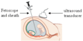
The procedure is usually performed under local anesthesia and may be done in an outpatient setting. The fetoscope is inserted through a small incision in the abdomen and into the uterus. The fetoscope allows the doctor to view the fetus, the umbilical cord, and the placenta. The procedure can be used to take biopsies of the placenta or the skin of the fetus, or to perform certain types of fetal surgery.
Uses[edit]
Fetoscopy is used for both diagnostic and therapeutic purposes. It can be used to diagnose certain genetic disorders and birth defects, and to perform procedures such as blood transfusions to the fetus, laser therapy for twin-to-twin transfusion syndrome, and fetal surgery for conditions such as spina bifida.
Risks[edit]
As with any surgical procedure, there are risks associated with fetoscopy. These may include preterm labor, infection, bleeding, and damage to the uterus or fetus. The risks should be discussed with the doctor before the procedure.
See also[edit]
References[edit]

This WikiMD article can only be edited by registered and verified editors. You can log in or register.
-
 Fetoscopy


Medical Disclaimer: WikiMD is for informational purposes only and is not a substitute for professional medical advice. Content may be inaccurate or outdated and should not be used for diagnosis or treatment. Always consult your healthcare provider for medical decisions. Verify information with trusted sources such as CDC.gov and NIH.gov. By using this site, you agree that WikiMD is not liable for any outcomes related to its content. See full disclaimer.
Credits:Most images are courtesy of Wikimedia commons, and templates, categories Wikipedia, licensed under CC BY SA or similar.
Translate page: - East Asian
中文,
日本,
한국어,
South Asian
हिन्दी,
தமிழ்,
తెలుగు,
Urdu,
ಕನ್ನಡ,
Southeast Asian
Indonesian,
Vietnamese,
Thai,
မြန်မာဘာသာ,
বাংলা
European
español,
Deutsch,
français,
Greek,
português do Brasil,
polski,
română,
русский,
Nederlands,
norsk,
svenska,
suomi,
Italian
Middle Eastern & African
عربى,
Turkish,
Persian,
Hebrew,
Afrikaans,
isiZulu,
Kiswahili,
Other
Bulgarian,
Hungarian,
Czech,
Swedish,
മലയാളം,
मराठी,
ਪੰਜਾਬੀ,
ગુજરાતી,
Portuguese,
Ukrainian
