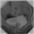
Cystography
Cystography is a diagnostic imaging test used to examine the structure and functionality of the bladder and the urethra. This procedure is particularly useful in identifying abnormalities such as bladder stones, tumors, diverticula, and vesicoureteral reflux. It involves the use of X-ray imaging and a contrast material to visualize the bladder's interior.
Procedure[edit]
Cystography is typically performed in a hospital's radiology department. The process begins with the patient lying on their back on an X-ray table. A healthcare professional then inserts a catheter into the urethra, through which a contrast dye is introduced into the bladder. The contrast dye makes the bladder area more visible on the X-ray images. Once the bladder is filled with the dye, the catheter is removed, and several X-rays are taken from different angles. In some cases, additional images may be taken while the patient urinates to assess the function of the bladder and urethra.
Types of Cystography[edit]
There are two main types of cystography:
- Retrograde Cystography: This is the most common type, where contrast dye is introduced into the bladder through a catheter.
- Voiding Cystourethrography (VCUG): This involves taking X-ray images while the patient is urinating to evaluate the bladder and urethra's function, particularly looking for vesicoureteral reflux.
Indications[edit]
Cystography is indicated for patients experiencing symptoms such as:
- Recurrent urinary tract infections
- Painful urination
- Unexplained hematuria (blood in the urine)
- Suspected bladder injury or trauma
- Evaluation of vesicoureteral reflux in children
Risks and Complications[edit]
While cystography is generally safe, there are potential risks and complications, including:
- Allergic reaction to the contrast dye
- Urinary tract infection
- Bladder injury
- Radiation exposure, though it is typically minimal
Preparation[edit]
Patients may be advised to drink plenty of fluids and possibly to take a mild laxative the day before the procedure to clear the bowel, which helps in obtaining clearer images. It is also important to inform the healthcare provider of any allergies, especially to iodine or contrast dyes, and of all medications being taken.
Aftercare[edit]
After the procedure, patients are encouraged to drink plenty of water to help flush the contrast dye from the system. Any signs of infection, such as fever, chills, or pain, should be reported to a healthcare provider immediately.
Conclusion[edit]
Cystography is a valuable diagnostic tool in the evaluation of bladder and urethral abnormalities. It provides critical information that can guide the management and treatment of various urological conditions. As with any medical procedure, patients should discuss the potential risks and benefits with their healthcare provider.
| Radiology | ||||||||||
|---|---|---|---|---|---|---|---|---|---|---|
This radiology-related article is a stub. You can help WikiMD by expanding it.
|
| Urology | ||||||||||
|---|---|---|---|---|---|---|---|---|---|---|
|
This medical article is a stub. You can help WikiMD by expanding the page. |
-
 Cystography


Medical Disclaimer: WikiMD is for informational purposes only and is not a substitute for professional medical advice. Content may be inaccurate or outdated and should not be used for diagnosis or treatment. Always consult your healthcare provider for medical decisions. Verify information with trusted sources such as CDC.gov and NIH.gov. By using this site, you agree that WikiMD is not liable for any outcomes related to its content. See full disclaimer.
Credits:Most images are courtesy of Wikimedia commons, and templates, categories Wikipedia, licensed under CC BY SA or similar.
Translate page: - East Asian
中文,
日本,
한국어,
South Asian
हिन्दी,
தமிழ்,
తెలుగు,
Urdu,
ಕನ್ನಡ,
Southeast Asian
Indonesian,
Vietnamese,
Thai,
မြန်မာဘာသာ,
বাংলা
European
español,
Deutsch,
français,
Greek,
português do Brasil,
polski,
română,
русский,
Nederlands,
norsk,
svenska,
suomi,
Italian
Middle Eastern & African
عربى,
Turkish,
Persian,
Hebrew,
Afrikaans,
isiZulu,
Kiswahili,
Other
Bulgarian,
Hungarian,
Czech,
Swedish,
മലയാളം,
मराठी,
ਪੰਜਾਬੀ,
ગુજરાતી,
Portuguese,
Ukrainian
