The Femoral Artery
Anatomy > Gray's Anatomy of the Human Body > VI. The Arteries > 6a. The Femoral Artery
Henry Gray (1821–1865). Anatomy of the Human Body. 1918.
The Femoral Artery
The femoral artery is a large artery in the thigh and the main arterial supply to the thigh and leg. It enters the thigh from behind the inguinal ligament as the continuation of the external iliac artery.
Here, it lies midway between the anterior superior iliac spine and the symphysis pubis. The femoral artery gives off the deep femoral artery or profunda femoris artery and descends along the anteromedial part of the thigh in the femoral triangle. It enters and passes through the adductor canal, and becomes the popliteal artery as it passes through the adductor hiatus in the adductor magnus near the junction of the middle and distal thirds of the thigh.<ref>,
 Thieme Atlas of Anatomy: General Anatomy and Musculoskeletal System,

 Thieme,
 2006,


 ISBN 978-3-13-142081-7,</ref>
Structure
Its first three or four centimetres are enclosed, with the femoral vein, in the femoral sheath.
Relations
The relations of the femoral artery are as follows:
- Anteriorly: In the upper part of its course, it is superficial and is covered by skin and fascia. In the lower part of its course, it passes behind the sartorius muscle.
- Posteriorly: The artery lies on the psoas, which separates it from the hip joint, the pectineus, and the adductor longus. The femoral vein intervenes between the artery and the adductor longus.
- Medially: It is related to the femoral vein in the upper part of its course.
- Laterally: The femoral nerve and its branches.
Branches

The femoral artery gives off several branches in the thigh which include;
- The superficial circumflex iliac artery is a small branch that runs up to the region of the anterior superior iliac spine.
- The superficial epigastric artery is a small branch that crosses the inguinal ligament and runs to the region of the umbilicus.
- The superficial external pudendal artery is a small branch that runs medially to supply the skin of the scrotum (or labium majus).
- The deep external pudendal artery runs medially and supplies the skin of the scrotum (or labium majus).
- The profunda femoris artery is a large and important branch that arises from the lateral side of the femoral artery about 1.5 in. (4 cm) below the inguinal ligament. It passes medially behind the femoral vessels and enters the medial fascial compartment of the thigh. It ends by becoming the fourth perforating artery. At its origin, it gives off the medial and lateral femoral circumflex arteries, and during its course it gives off three perforating arteries.
- The descending genicular artery is a small branch that arises from the femoral artery near its termination within the adductor canal. It assists in supplying the knee joint.
Segments

In clinical parlance, the femoral artery has the following segments:
- The common femoral artery is the segment of the femoral artery between the inferior margin of the inguinal ligament and the branching point of the deep femoral artery.
- The subsartorial artery<ref name=subsartorial>Mikael Häggström,
 Subsartorial Vessels as Replacement Name for Superficial Femoral Vessels,
 International Journal of Anatomy, Radiology and Surgery,
 2019,

 pp. AV01–AV02,


 [Ra1_F(SHU)_PF1(A_SHU)_PFA(A_SHU)_PF2(AKA_SHU)_PN(SHU).pdf Full text],</ref> or superficial femoral artery<ref>Richard S.,

 Clinical Anatomy By Regions. online version,
 8 edition,
 Baltimore:Lippincott Williams & Wilkins,
 2008,


 ISBN 978-0-7817-6404-9,


 Pages: 581–582,</ref> are designations for the segment between the branching point of the deep femoral vein and the adductor hiatus, passing through the subsartorial canal. However, usage of the term superficial femoral is discouraged by many physicians because it leads to confusion among general medical practitioners, at least for the femoral vein that courses next to the femoral artery.<ref name=bundens_7563535>,
 The superficial femoral vein. A potentially lethal misnomer,
 JAMA,
 1995,
 Vol. 274(Issue: 16),
 pp. 1296–8,
 DOI: 10.1001/jama.1995.03530160048032,
 PMID: 7563535,</ref> In particular, the adjacent femoral vein is clinically a deep vein, where deep vein thrombosis indicates anticoagulant or thrombolytic therapy, but the adjective "superficial" leads many physicians to falsely believe it is a superficial vein, which has resulted in patients with femoral thrombosis being denied proper treatment.<ref name=hammond_14595157>,
 The superficial femoral vein,
 Radiology,
 2003,
 Vol. 229(Issue: 2),

 DOI: 10.1148/radiol.2292030418,
 PMID: 14595157,</ref><ref name="pmid20980677">Kitchens CS,
 How I treat superficial venous thrombosis.,
 Blood,
 2011,
 Vol. 117(Issue: 1),
 pp. 39–44,
 DOI: 10.1182/blood-2010-05-286690,
 PMID: 20980677,</ref><ref>,
 Use of the term "superficial femoral vein" in ultrasound.,
 J Clin Ultrasound,
 2011,
 Vol. 39(Issue: 1),
 pp. 32–34,
 DOI: 10.1002/jcu.20747,
 PMID: 20957733,</ref> Therefore, the terms subsartorial artery and subsartorial vein have been suggested for the femoral artery and vein, respectively, distally to the branching points of the deep femoral artery and vein.<ref name=subsartorial/>
Additional images
-
 Structures passing behind the inguinal ligament. (Femoral artery labeled at upper right.)
 -
 Cross-section showing structures surrounding right hip-joint.
 -
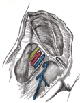 Femoral sheath laid open to show its three compartments.
 -
 The femoral artery.
 -
 The spermatic cord in the inguinal canal.
 -
 Front of right thigh, showing surface markings for bones, femoral artery and femoral nerve.
 -
 Femoral artery and its major branches - right thigh, anterior view.
 -
 Illustration depicting main leg arteries (anterior view).
 -
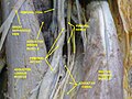 Femoral artery - deep dissection.
 -
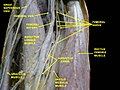 Femoral artery - deep dissection.


External links
- Anatomy photo:12:05-0101 at the SUNY Downstate Medical Center
- Template:ViennaCrossSection
- Image at umich.edu - pulse
- Diagram at MSU
- QuantaFlo vs ABI in Peripheral Arterial Disease
| Arteries of the human leg | ||||||||||||||||||||||||||||
|---|---|---|---|---|---|---|---|---|---|---|---|---|---|---|---|---|---|---|---|---|---|---|---|---|---|---|---|---|
|
Gray's Anatomy
- Gray's Anatomy Contents
- Gray's Anatomy Subject Index
- About Classic Gray's Anatomy
- Glossary of anatomy terms
Anatomy atlases (external)
[1] - Anatomy Atlases
 | |
|---|---|
|
|
 |
| Human systems and organs | ||||||||||||||
|---|---|---|---|---|---|---|---|---|---|---|---|---|---|---|
|
Ad. Transform your life with W8MD's
GLP-1 weight loss injections special from $29.99 with insurance
|
WikiMD Medical Encyclopedia |
Medical Disclaimer: WikiMD is for informational purposes only and is not a substitute for professional medical advice. Content may be inaccurate or outdated and should not be used for diagnosis or treatment. Always consult your healthcare provider for medical decisions. Verify information with trusted sources such as CDC.gov and NIH.gov. By using this site, you agree that WikiMD is not liable for any outcomes related to its content. See full disclaimer.
Credits:Most images are courtesy of Wikimedia commons, and templates, categories Wikipedia, licensed under CC BY SA or similar.
Translate this page: - East Asian
中文,
日本,
한국어,
South Asian
हिन्दी,
தமிழ்,
తెలుగు,
Urdu,
ಕನ್ನಡ,
Southeast Asian
Indonesian,
Vietnamese,
Thai,
မြန်မာဘာသာ,
বাংলা
European
español,
Deutsch,
français,
Greek,
português do Brasil,
polski,
română,
русский,
Nederlands,
norsk,
svenska,
suomi,
Italian
Middle Eastern & African
عربى,
Turkish,
Persian,
Hebrew,
Afrikaans,
isiZulu,
Kiswahili,
Other
Bulgarian,
Hungarian,
Czech,
Swedish,
മലയാളം,
मराठी,
ਪੰਜਾਬੀ,
ગુજરાતી,
Portuguese,
Ukrainian
