Femoral triangle
The Femoral Triangle (also known as Scarpa's triangle) is an anatomical region of the upper inner human thigh. It is a subfascial space which in living people appears as a triangular depression inferior to the inguinal ligament when the thigh is flexed, abducted and laterally rotated.
Etymology[edit]
The term "Femoral" refers to the femur or the thigh bone, the largest bone in the human body. The term "Triangle" is used to describe the shape of this anatomical region. The term "Scarpa's triangle" is named after Antonio Scarpa, an Italian anatomist.
Boundaries[edit]
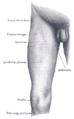
The femoral triangle is bounded:
- Superiorly: by the Inguinal Ligament
- Medially: by the medial border of the Adductor Longus Muscle
- Laterally: by the medial border of the Sartorius Muscle
The floor of the femoral triangle is formed by the Pectineus and Adductor Longus muscles medially and the Iliacus and Psoas Major muscles laterally. The roof of the femoral triangle is formed by skin, superficial and deep fascia.
Contents[edit]
The femoral triangle contains (from lateral to medial):
- The Femoral Nerve
- The Femoral Artery
- The Femoral Vein
- The Deep Inguinal Lymph Nodes
Clinical Significance[edit]
The femoral triangle is important in medicine and surgery, particularly for surgical access to the femoral artery and femoral vein. It is also a landmark for the inguinal lymph nodes, which can be palpated during a physical examination to assess for lymphadenopathy.
See Also[edit]
- Femoral Artery
- Femoral Vein
- Femoral Nerve
- Inguinal Ligament
- Adductor Longus Muscle
- Sartorius Muscle
- Pectineus
- Iliacus
- Psoas Major
- Deep Inguinal Lymph Nodes

This WikiMD article can only be edited by registered and verified editors. You can log in or register.
-
3D Tour of the Femoral Triangle
-
 Gray's Anatomy illustration of the Femoral Triangle
 -
Borders of the Femoral Triangle
-
Contents of the Femoral Triangle


Medical Disclaimer: WikiMD is for informational purposes only and is not a substitute for professional medical advice. Content may be inaccurate or outdated and should not be used for diagnosis or treatment. Always consult your healthcare provider for medical decisions. Verify information with trusted sources such as CDC.gov and NIH.gov. By using this site, you agree that WikiMD is not liable for any outcomes related to its content. See full disclaimer.
Credits:Most images are courtesy of Wikimedia commons, and templates, categories Wikipedia, licensed under CC BY SA or similar.
Translate page: - East Asian
中文,
日本,
한국어,
South Asian
हिन्दी,
தமிழ்,
తెలుగు,
Urdu,
ಕನ್ನಡ,
Southeast Asian
Indonesian,
Vietnamese,
Thai,
မြန်မာဘာသာ,
বাংলা
European
español,
Deutsch,
français,
Greek,
português do Brasil,
polski,
română,
русский,
Nederlands,
norsk,
svenska,
suomi,
Italian
Middle Eastern & African
عربى,
Turkish,
Persian,
Hebrew,
Afrikaans,
isiZulu,
Kiswahili,
Other
Bulgarian,
Hungarian,
Czech,
Swedish,
മലയാളം,
मराठी,
ਪੰਜਾਬੀ,
ગુજરાતી,
Portuguese,
Ukrainian
