Accessory spleen
Accessory Spleen
An accessory spleen is a small nodule of splenic tissue that is separate from the main body of the spleen. It is a common congenital anomaly, occurring in approximately 10-30% of the population. Accessory spleens are usually asymptomatic and are often discovered incidentally during imaging studies or surgical procedures.
Anatomy and Location
Accessory spleens are typically found near the hilum of the spleen, but they can also be located in other areas of the abdominal cavity, such as the pancreas, omentum, or mesentery. They are usually small, ranging from a few millimeters to a few centimeters in diameter.
Development
Accessory spleens develop during embryogenesis when splenic tissue becomes separated from the main body of the spleen. This can occur due to incomplete fusion of the splenic anlage or due to the presence of multiple splenic nodules during development.
Clinical Significance
While accessory spleens are generally benign and asymptomatic, they can become clinically significant in certain situations. For example, in patients undergoing splenectomy for conditions such as idiopathic thrombocytopenic purpura (ITP), an accessory spleen can continue to produce platelets, potentially leading to a recurrence of symptoms. In such cases, identification and removal of the accessory spleen may be necessary.
Diagnosis
Accessory spleens are often identified through imaging studies such as computed tomography (CT) scans, magnetic resonance imaging (MRI), or ultrasound. On imaging, they appear as well-defined, homogeneous nodules with similar characteristics to the main spleen.
Treatment
In most cases, accessory spleens do not require treatment. However, if they cause symptoms or complications, surgical removal may be indicated. This is typically done laparoscopically.
Related Pages
Gallery
-
 CT scan of an accessory spleen
 -
 Gross pathology of an accessory spleen
 -
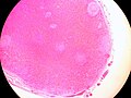 Photomicrograph of a splenunculus
 -
 Accessory spleen (Nebenmilz)
 -
 Hypertrophic accessory spleen with hematoma


_photomicrograph.JPG)


Accessory spleen
-
CT scan of an accessory spleen
-
Gross pathology of an accessory spleen
-
Splenunculus (accessory spleen) photomicrograph
-
Accessory spleen
-
Hypertrophic accessory spleen with hematoma
Ad. Transform your life with W8MD's
GLP-1 weight loss injections special from $29.99 with insurance
|
WikiMD Medical Encyclopedia |
Medical Disclaimer: WikiMD is for informational purposes only and is not a substitute for professional medical advice. Content may be inaccurate or outdated and should not be used for diagnosis or treatment. Always consult your healthcare provider for medical decisions. Verify information with trusted sources such as CDC.gov and NIH.gov. By using this site, you agree that WikiMD is not liable for any outcomes related to its content. See full disclaimer.
Credits:Most images are courtesy of Wikimedia commons, and templates, categories Wikipedia, licensed under CC BY SA or similar.
Translate this page: - East Asian
中文,
日本,
한국어,
South Asian
हिन्दी,
தமிழ்,
తెలుగు,
Urdu,
ಕನ್ನಡ,
Southeast Asian
Indonesian,
Vietnamese,
Thai,
မြန်မာဘာသာ,
বাংলা
European
español,
Deutsch,
français,
Greek,
português do Brasil,
polski,
română,
русский,
Nederlands,
norsk,
svenska,
suomi,
Italian
Middle Eastern & African
عربى,
Turkish,
Persian,
Hebrew,
Afrikaans,
isiZulu,
Kiswahili,
Other
Bulgarian,
Hungarian,
Czech,
Swedish,
മലയാളം,
मराठी,
ਪੰਜਾਬੀ,
ગુજરાતી,
Portuguese,
Ukrainian
