Arecoline
Medical imaging technique
Aortography is a medical imaging technique used to visualize the aorta, the largest artery in the human body. This procedure is a type of angiography, which involves the use of contrast media to enhance the visibility of blood vessels on X-ray images. Aortography is primarily used to diagnose and evaluate conditions affecting the aorta, such as aortic aneurysm, aortic dissection, and aortic stenosis.
Procedure
Aortography is typically performed in a catheterization laboratory. The procedure begins with the insertion of a catheter into a large artery, usually the femoral artery in the groin. The catheter is then guided through the arterial system to the aorta. Once in place, a contrast agent is injected through the catheter, and X-ray images are taken to visualize the aorta and its branches.
The procedure can be performed using different techniques, including:
- Digital Subtraction Angiography (DSA): This technique involves taking two sets of images, one before and one after the injection of contrast. The pre-contrast images are subtracted from the post-contrast images to enhance the visibility of the blood vessels.
- Computed Tomography Angiography (CTA): This is a non-invasive alternative that uses CT scanning to obtain detailed images of the aorta after the injection of contrast material.
- Magnetic Resonance Angiography (MRA): This technique uses magnetic resonance imaging to visualize the aorta without the need for ionizing radiation.
Indications
Aortography is indicated for the evaluation of various aortic pathologies, including:
- Aortic aneurysm: Aortography can help determine the size and extent of an aneurysm.
- Aortic dissection: The procedure can identify the presence and location of a dissection.
- Aortic stenosis: Aortography can assess the severity of stenosis and its impact on blood flow.
- Congenital heart defects: It can be used to evaluate structural abnormalities of the aorta.
Risks and Complications
As with any invasive procedure, aortography carries certain risks, including:
- Allergic reaction to the contrast material
- Bleeding or hematoma at the catheter insertion site
- Infection
- Kidney damage due to contrast material
- Radiation exposure
History
The development of aortography dates back to the early 20th century, with significant advancements in imaging technology and techniques over the decades. The introduction of digital subtraction angiography in the 1970s revolutionized the field by providing clearer images with less contrast material.
Related pages
Gallery
-
 Aortography image showing calcification in the aorta.

-
 Arecoline chemical structure
 -
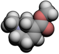 3D model of Arecoline
 -
 Synthesis pathway of Arecoline
 -
 Alternative synthesis of Arecoline


Ad. Transform your life with W8MD's Budget GLP-1 injections from $75


W8MD offers a medical weight loss program to lose weight in Philadelphia. Our physician-supervised medical weight loss provides:
- Weight loss injections in NYC (generic and brand names):
- Zepbound / Mounjaro, Wegovy / Ozempic, Saxenda
- Most insurances accepted or discounted self-pay rates. We will obtain insurance prior authorizations if needed.
- Generic GLP1 weight loss injections from $75 for the starting dose.
- Also offer prescription weight loss medications including Phentermine, Qsymia, Diethylpropion, Contrave etc.
NYC weight loss doctor appointmentsNYC weight loss doctor appointments
Start your NYC weight loss journey today at our NYC medical weight loss and Philadelphia medical weight loss clinics.
- Call 718-946-5500 to lose weight in NYC or for medical weight loss in Philadelphia 215-676-2334.
- Tags:NYC medical weight loss, Philadelphia lose weight Zepbound NYC, Budget GLP1 weight loss injections, Wegovy Philadelphia, Wegovy NYC, Philadelphia medical weight loss, Brookly weight loss and Wegovy NYC
![]()
![]()
![]()
![]()
|
WikiMD's Wellness Encyclopedia |
| Let Food Be Thy Medicine Medicine Thy Food - Hippocrates |
Medical Disclaimer: WikiMD is not a substitute for professional medical advice. The information on WikiMD is provided as an information resource only, may be incorrect, outdated or misleading, and is not to be used or relied on for any diagnostic or treatment purposes. Please consult your health care provider before making any healthcare decisions or for guidance about a specific medical condition. WikiMD expressly disclaims responsibility, and shall have no liability, for any damages, loss, injury, or liability whatsoever suffered as a result of your reliance on the information contained in this site. By visiting this site you agree to the foregoing terms and conditions, which may from time to time be changed or supplemented by WikiMD. If you do not agree to the foregoing terms and conditions, you should not enter or use this site. See full disclaimer.
Credits:Most images are courtesy of Wikimedia commons, and templates, categories Wikipedia, licensed under CC BY SA or similar.
Translate this page: - East Asian
中文,
日本,
한국어,
South Asian
हिन्दी,
தமிழ்,
తెలుగు,
Urdu,
ಕನ್ನಡ,
Southeast Asian
Indonesian,
Vietnamese,
Thai,
မြန်မာဘာသာ,
বাংলা
European
español,
Deutsch,
français,
Greek,
português do Brasil,
polski,
română,
русский,
Nederlands,
norsk,
svenska,
suomi,
Italian
Middle Eastern & African
عربى,
Turkish,
Persian,
Hebrew,
Afrikaans,
isiZulu,
Kiswahili,
Other
Bulgarian,
Hungarian,
Czech,
Swedish,
മലയാളം,
मराठी,
ਪੰਜਾਬੀ,
ગુજરાતી,
Portuguese,
Ukrainian
