Aneurysmal bone cyst: Difference between revisions
CSV import |
CSV import |
||
| Line 4: | Line 4: | ||
File:Aneurysmal_bone_cyst_-_intermed_mag.jpg|Aneurysmal bone cyst - intermediate magnification | File:Aneurysmal_bone_cyst_-_intermed_mag.jpg|Aneurysmal bone cyst - intermediate magnification | ||
</gallery> | </gallery> | ||
== Aneurysmal Bone Cyst == | |||
An '''aneurysmal bone cyst''' (ABC) is a benign, blood-filled bone lesion that can cause significant bone destruction and deformity. It is characterized by the presence of blood-filled cystic spaces separated by connective tissue septa containing osteoclast-type giant cells, fibroblasts, and reactive woven bone. | |||
== Pathophysiology == | |||
The exact cause of aneurysmal bone cysts is not well understood. However, it is believed that they may arise due to a vascular malformation or a response to a preceding bone lesion, such as a [[giant cell tumor of bone]], [[chondroblastoma]], or [[fibrous dysplasia]]. The cysts are expansile and can cause thinning of the surrounding bone cortex, leading to potential fracture or deformity. | |||
== Clinical Presentation == | |||
Patients with aneurysmal bone cysts typically present with localized pain and swelling. The lesions are most commonly found in the [[metaphysis]] of long bones, such as the [[femur]], [[tibia]], and [[humerus]], but can also occur in the [[vertebrae]] and [[pelvis]]. The condition is most frequently diagnosed in individuals under the age of 20. | |||
== Diagnosis == | |||
Diagnosis of an aneurysmal bone cyst is primarily based on imaging studies. [[X-ray]] imaging typically shows an expansile, lytic lesion with a "blow-out" appearance. [[Magnetic resonance imaging]] (MRI) and [[computed tomography]] (CT) scans can provide more detailed information about the lesion's extent and its effect on surrounding structures. A definitive diagnosis is often confirmed through a [[biopsy]], which reveals the characteristic blood-filled cystic spaces and septa. | |||
== Treatment == | |||
The treatment of aneurysmal bone cysts often involves surgical intervention. The most common surgical procedure is curettage, where the cyst is scraped out of the bone. This may be followed by bone grafting to fill the defect. In some cases, more extensive surgery, such as en bloc resection, may be necessary. Adjuvant therapies, such as cryotherapy or sclerotherapy, may be used to reduce the risk of recurrence. | |||
== Prognosis == | |||
The prognosis for patients with aneurysmal bone cysts is generally good, especially with appropriate treatment. However, there is a risk of recurrence, particularly if the lesion is not completely removed. Recurrence rates can be as high as 20-30% in some cases. | |||
== Related Pages == | |||
* [[Bone tumor]] | |||
* [[Giant cell tumor of bone]] | |||
* [[Chondroblastoma]] | |||
* [[Fibrous dysplasia]] | |||
* [[Osteosarcoma]] | |||
{{Bone diseases}} | |||
[[Category:Bone disorders]] | |||
[[Category:Benign neoplasms]] | |||
Revision as of 00:40, 19 February 2025
-
 Aneurysmal bone cyst - very high magnification
 -
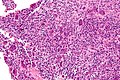 Aneurysmal bone cyst - high magnification
 -
 Aneurysmal bone cyst - intermediate magnification


Aneurysmal Bone Cyst
An aneurysmal bone cyst (ABC) is a benign, blood-filled bone lesion that can cause significant bone destruction and deformity. It is characterized by the presence of blood-filled cystic spaces separated by connective tissue septa containing osteoclast-type giant cells, fibroblasts, and reactive woven bone.
Pathophysiology
The exact cause of aneurysmal bone cysts is not well understood. However, it is believed that they may arise due to a vascular malformation or a response to a preceding bone lesion, such as a giant cell tumor of bone, chondroblastoma, or fibrous dysplasia. The cysts are expansile and can cause thinning of the surrounding bone cortex, leading to potential fracture or deformity.
Clinical Presentation
Patients with aneurysmal bone cysts typically present with localized pain and swelling. The lesions are most commonly found in the metaphysis of long bones, such as the femur, tibia, and humerus, but can also occur in the vertebrae and pelvis. The condition is most frequently diagnosed in individuals under the age of 20.
Diagnosis
Diagnosis of an aneurysmal bone cyst is primarily based on imaging studies. X-ray imaging typically shows an expansile, lytic lesion with a "blow-out" appearance. Magnetic resonance imaging (MRI) and computed tomography (CT) scans can provide more detailed information about the lesion's extent and its effect on surrounding structures. A definitive diagnosis is often confirmed through a biopsy, which reveals the characteristic blood-filled cystic spaces and septa.
Treatment
The treatment of aneurysmal bone cysts often involves surgical intervention. The most common surgical procedure is curettage, where the cyst is scraped out of the bone. This may be followed by bone grafting to fill the defect. In some cases, more extensive surgery, such as en bloc resection, may be necessary. Adjuvant therapies, such as cryotherapy or sclerotherapy, may be used to reduce the risk of recurrence.
Prognosis
The prognosis for patients with aneurysmal bone cysts is generally good, especially with appropriate treatment. However, there is a risk of recurrence, particularly if the lesion is not completely removed. Recurrence rates can be as high as 20-30% in some cases.
Related Pages
| Bone diseases | ||||||||||
|---|---|---|---|---|---|---|---|---|---|---|
This bone disease related article is a stub.
|
