Negri body: Difference between revisions
CSV import Tags: mobile edit mobile web edit |
CSV import |
||
| Line 26: | Line 26: | ||
[[Category:Infectious diseases]] | [[Category:Infectious diseases]] | ||
{{medicine-stub}} | {{medicine-stub}} | ||
<gallery> | |||
File:Histopathology_of_Negri_bodies_in_rabies_encephalitis.png|Histopathology of Negri bodies in rabies encephalitis | |||
File:Rabies_Virus_EM_PHIL_1876.JPG|Negri body | |||
</gallery> | |||
Latest revision as of 01:58, 18 February 2025
Negri bodies are distinct, eosinophilic, cytoplasmic inclusions predominantly found in the neurons of animals and humans infected with the Rabies virus, a member of the Rymoviridae family. First described by and named after Adelchi Negri in 1903, these inclusions are a hallmark of rabies infection and serve as an important diagnostic feature in post-mortem examination.
Characteristics[edit]
Negri bodies vary in size, typically ranging from 0.25 to 27 micrometers in diameter. They are most commonly observed in the Pyramidal cells of the Hippocampus, Purkinje cells of the Cerebellum, and neurons of the Cerebral cortex. Morphologically, Negri bodies appear as round or oval structures with a basophilic core surrounded by a clear halo and an outer eosinophilic ring when stained with Eosin and hematoxylin.
Pathogenesis[edit]
The exact function and composition of Negri bodies are not fully understood, but they are believed to be sites of viral replication and assembly. The presence of Rabies virus nucleoprotein and other viral components within Negri bodies has been confirmed through immunohistochemistry and electron microscopy. This suggests that Negri bodies play a crucial role in the rabies virus life cycle, facilitating the accumulation and assembly of viral particles.
Diagnosis[edit]
The detection of Negri bodies in brain tissue is a classical method for diagnosing rabies post-mortem. Techniques such as Direct fluorescent antibody test (dFA) and polymerase chain reaction (PCR) have surpassed the use of Negri body identification in terms of sensitivity and specificity. However, in resource-limited settings, the observation of Negri bodies in a brain smear stained with Sellers' stain remains a practical diagnostic approach.
Clinical Significance[edit]
The identification of Negri bodies in the brain tissue of an individual or animal with neurological symptoms suggestive of rabies provides a definitive diagnosis of the disease. Given the fatal nature of rabies once clinical symptoms appear, early detection and confirmation of the virus through the presence of Negri bodies or other diagnostic means are critical for public health measures and the initiation of post-exposure prophylaxis in potential human contacts.
Prevention and Control[edit]
Prevention of rabies is primarily through vaccination of domestic animals, wildlife control, and post-exposure prophylaxis in humans following potential exposure to the virus. Education on avoiding contact with wild animals and the importance of vaccinating pets are also key components of rabies control programs.
See Also[edit]

-
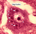 Histopathology of Negri bodies in rabies encephalitis
 -
 Negri body


