Mueller–Weiss syndrome: Difference between revisions
CSV import |
CSV import |
||
| Line 1: | Line 1: | ||
{{SI}} | |||
{{Infobox medical condition | |||
| name = Mueller–Weiss syndrome | |||
| image = [[File:Tarsus2.png|thumb|Illustration of the tarsal bones of the foot]] | |||
| caption = The tarsal bones of the foot, with the navicular bone highlighted | |||
| synonyms = Brailsford disease | |||
| pronunciation = | |||
| specialty = [[Orthopedics]], [[Podiatry]] | |||
| symptoms = [[Foot pain]], [[midfoot pain]], [[arch pain]] | |||
| onset = Typically in [[adulthood]] | |||
| duration = Chronic | |||
| causes = Unknown, possibly [[vascular insufficiency]] | |||
| risks = [[Female gender]], [[obesity]], [[trauma]] | |||
| diagnosis = [[Clinical examination]], [[X-ray]], [[MRI]] | |||
| differential = [[Osteoarthritis]], [[stress fracture]], [[Köhler disease]] | |||
| prevention = None known | |||
| treatment = [[Orthotics]], [[pain management]], [[surgery]] | |||
| prognosis = Variable, can lead to [[chronic pain]] | |||
| frequency = Rare | |||
}} | |||
'''Mueller–Weiss syndrome''' (MWS), also known as '''Spontaneous Osteonecrosis of the Tarsal Navicular in Adults''', is a rare, spontaneous [[osteonecrosis]] of the [[tarsal navicular]] in adults. It was first described by Mueller and Weiss in 1927. | '''Mueller–Weiss syndrome''' (MWS), also known as '''Spontaneous Osteonecrosis of the Tarsal Navicular in Adults''', is a rare, spontaneous [[osteonecrosis]] of the [[tarsal navicular]] in adults. It was first described by Mueller and Weiss in 1927. | ||
==Etiology== | ==Etiology== | ||
The exact cause of Mueller–Weiss syndrome is unknown. However, it is believed to be due to a combination of biomechanical and vascular factors. The navicular bone is subjected to high compressive forces, which may lead to stress fractures and subsequent osteonecrosis. Additionally, the blood supply to the navicular bone is precarious, which may contribute to the development of osteonecrosis. | The exact cause of Mueller–Weiss syndrome is unknown. However, it is believed to be due to a combination of biomechanical and vascular factors. The navicular bone is subjected to high compressive forces, which may lead to stress fractures and subsequent osteonecrosis. Additionally, the blood supply to the navicular bone is precarious, which may contribute to the development of osteonecrosis. | ||
==Clinical Presentation== | ==Clinical Presentation== | ||
Patients with Mueller–Weiss syndrome typically present with chronic, midfoot pain that is exacerbated by weight-bearing activities. Physical examination may reveal swelling and tenderness over the navicular bone. | Patients with Mueller–Weiss syndrome typically present with chronic, midfoot pain that is exacerbated by weight-bearing activities. Physical examination may reveal swelling and tenderness over the navicular bone. | ||
==Diagnosis== | ==Diagnosis== | ||
The diagnosis of Mueller–Weiss syndrome is primarily based on clinical presentation and imaging studies. [[Radiography]] is typically the first imaging modality used. However, [[Magnetic Resonance Imaging|MRI]] is more sensitive and specific in detecting early changes of osteonecrosis. | The diagnosis of Mueller–Weiss syndrome is primarily based on clinical presentation and imaging studies. [[Radiography]] is typically the first imaging modality used. However, [[Magnetic Resonance Imaging|MRI]] is more sensitive and specific in detecting early changes of osteonecrosis. | ||
==Treatment== | ==Treatment== | ||
The treatment of Mueller–Weiss syndrome is primarily conservative and includes rest, [[Nonsteroidal anti-inflammatory drug|NSAIDs]], and orthotic devices. In refractory cases, surgical intervention may be required. | The treatment of Mueller–Weiss syndrome is primarily conservative and includes rest, [[Nonsteroidal anti-inflammatory drug|NSAIDs]], and orthotic devices. In refractory cases, surgical intervention may be required. | ||
==Prognosis== | ==Prognosis== | ||
The prognosis of Mueller–Weiss syndrome is variable. Some patients may experience resolution of symptoms with conservative treatment, while others may require surgical intervention. Long-term outcomes are generally favorable with appropriate treatment. | The prognosis of Mueller–Weiss syndrome is variable. Some patients may experience resolution of symptoms with conservative treatment, while others may require surgical intervention. Long-term outcomes are generally favorable with appropriate treatment. | ||
==Gallery== | |||
<gallery> | |||
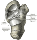
File:Navicular_bone_-_posterior_view.jpg|Posterior view of the navicular bone | |||
File:Gray359.png|Anatomical illustration of the foot bones | |||
</gallery> | |||
==See Also== | ==See Also== | ||
* [[Osteonecrosis]] | * [[Osteonecrosis]] | ||
| Line 26: | Line 40: | ||
* [[Radiography]] | * [[Radiography]] | ||
* [[Magnetic Resonance Imaging]] | * [[Magnetic Resonance Imaging]] | ||
[[Category:Orthopedic disorders]] | [[Category:Orthopedic disorders]] | ||
[[Category:Rare diseases]] | [[Category:Rare diseases]] | ||
| Line 33: | Line 46: | ||
{{Orthopedic conditions}} | {{Orthopedic conditions}} | ||
[[Category:Syndromes]] {{stub}} | [[Category:Syndromes]] {{stub}} | ||
Latest revision as of 04:46, 6 April 2025

Editor-In-Chief: Prab R Tumpati, MD
Obesity, Sleep & Internal medicine
Founder, WikiMD Wellnesspedia &
W8MD medical weight loss NYC and sleep center NYC
| Mueller–Weiss syndrome | |
|---|---|
 | |
| Synonyms | Brailsford disease |
| Pronounce | N/A |
| Specialty | Orthopedics, Podiatry |
| Symptoms | Foot pain, midfoot pain, arch pain |
| Complications | N/A |
| Onset | Typically in adulthood |
| Duration | Chronic |
| Types | N/A |
| Causes | Unknown, possibly vascular insufficiency |
| Risks | Female gender, obesity, trauma |
| Diagnosis | Clinical examination, X-ray, MRI |
| Differential diagnosis | Osteoarthritis, stress fracture, Köhler disease |
| Prevention | None known |
| Treatment | Orthotics, pain management, surgery |
| Medication | N/A |
| Prognosis | Variable, can lead to chronic pain |
| Frequency | Rare |
| Deaths | N/A |
Mueller–Weiss syndrome (MWS), also known as Spontaneous Osteonecrosis of the Tarsal Navicular in Adults, is a rare, spontaneous osteonecrosis of the tarsal navicular in adults. It was first described by Mueller and Weiss in 1927.
Etiology[edit]
The exact cause of Mueller–Weiss syndrome is unknown. However, it is believed to be due to a combination of biomechanical and vascular factors. The navicular bone is subjected to high compressive forces, which may lead to stress fractures and subsequent osteonecrosis. Additionally, the blood supply to the navicular bone is precarious, which may contribute to the development of osteonecrosis.
Clinical Presentation[edit]
Patients with Mueller–Weiss syndrome typically present with chronic, midfoot pain that is exacerbated by weight-bearing activities. Physical examination may reveal swelling and tenderness over the navicular bone.
Diagnosis[edit]
The diagnosis of Mueller–Weiss syndrome is primarily based on clinical presentation and imaging studies. Radiography is typically the first imaging modality used. However, MRI is more sensitive and specific in detecting early changes of osteonecrosis.
Treatment[edit]
The treatment of Mueller–Weiss syndrome is primarily conservative and includes rest, NSAIDs, and orthotic devices. In refractory cases, surgical intervention may be required.
Prognosis[edit]
The prognosis of Mueller–Weiss syndrome is variable. Some patients may experience resolution of symptoms with conservative treatment, while others may require surgical intervention. Long-term outcomes are generally favorable with appropriate treatment.
Gallery[edit]
-
 Posterior view of the navicular bone
 -
 Anatomical illustration of the foot bones


See Also[edit]
NIH genetic and rare disease info[edit]
Mueller–Weiss syndrome is a rare disease.
| Rare and genetic diseases | ||||||
|---|---|---|---|---|---|---|
|
Rare diseases - Mueller–Weiss syndrome
|
| Orthopedic Conditions | ||||||||||
|---|---|---|---|---|---|---|---|---|---|---|
This orthopedic conditions related article is a stub.
|


