Fetal thrombotic vasculopathy: Difference between revisions
CSV import |
CSV import Tags: mobile edit mobile web edit |
||
| Line 21: | Line 21: | ||
{{medicine-stub}} | {{medicine-stub}} | ||
<gallery> | |||
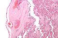
File:Fetal_thrombotic_vasculopathy_-_intermed_mag.jpg|Fetal thrombotic vasculopathy - intermediate magnification | |||
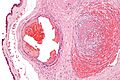
File:Fetal_thrombotic_vasculopathy_-_low_mag.jpg|Fetal thrombotic vasculopathy - low magnification | |||
File:Fetal_thrombotic_vasculopathy_-_high_mag.jpg|Fetal thrombotic vasculopathy - high magnification | |||
File:Fetal_thrombotic_vasculopathy_-_very_high_mag.jpg|Fetal thrombotic vasculopathy - very high magnification | |||
</gallery> | |||
Revision as of 23:57, 24 February 2025


Fetal Thrombotic Vasculopathy (FTV) is a pathological condition affecting the placenta that can have significant implications for fetal development and outcomes. It is characterized by the presence of thrombosis (blood clots) within the fetal vessels of the placenta, leading to compromised blood flow and oxygen delivery to the fetus. This condition is associated with a range of adverse fetal outcomes, including intrauterine growth restriction (IUGR), fetal distress, and in severe cases, stillbirth.
Etiology
The exact cause of Fetal Thrombotic Vasculopathy is not fully understood, but it is believed to be multifactorial. Factors that may contribute to the development of FTV include maternal conditions such as diabetes mellitus, hypertension, and antiphospholipid syndrome, as well as fetal factors like genetic thrombophilias. Infections and placental insufficiency have also been implicated in the pathogenesis of FTV.
Pathophysiology
FTV involves the formation of blood clots within the small blood vessels of the placenta, particularly affecting the villous capillaries and stem vessels. This leads to reduced blood flow and oxygen exchange between the maternal and fetal circulations. The resultant hypoxia and ischemia can cause necrosis of placental tissue and impact fetal development. The condition can be focal, affecting limited areas of the placenta, or widespread, leading to significant placental dysfunction.
Clinical Features and Diagnosis
FTV is often asymptomatic and may only be diagnosed postnatally through histopathological examination of the placenta. In some cases, signs of fetal distress or IUGR may prompt further investigation. Ultrasound and Doppler studies can suggest placental insufficiency, but definitive diagnosis requires placental pathology.
Management and Prognosis
Management of Fetal Thrombotic Vasculopathy primarily focuses on monitoring and addressing any underlying maternal conditions. Close surveillance of fetal well-being through non-stress tests, biophysical profiles, and Doppler ultrasound assessments is crucial. In severe cases, early delivery may be considered to prevent adverse outcomes. The prognosis of FTV depends on the extent of placental involvement and the presence of any underlying maternal or fetal conditions. Early detection and management are key to improving outcomes.
Prevention
Preventive strategies for Fetal Thrombotic Vasculopathy include optimal management of maternal health conditions, such as control of diabetes and hypertension, and treatment of any identified thrombophilias with anticoagulation therapy. Prenatal care and regular monitoring of high-risk pregnancies are essential for early detection and intervention.

-
 Fetal thrombotic vasculopathy - intermediate magnification
 -
 Fetal thrombotic vasculopathy - low magnification
 -
 Fetal thrombotic vasculopathy - high magnification
 -
 Fetal thrombotic vasculopathy - very high magnification

