Myelography
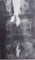
Myelography is a type of radiographic examination that uses a contrast medium to detect pathology of the spinal cord, including the location of a spinal cord injury, cysts, and tumors. This procedure is typically performed by a radiologist and uses X-ray imaging or computed tomography (CT) scanning.
Procedure[edit]
The procedure involves the injection of a contrast medium into the subarachnoid space in the spine. This is usually done in the lower back where the cauda equina of the spinal cord is located. The patient is then tilted in various directions to distribute the contrast medium and improve the images. The procedure is usually followed by a CT scan to further enhance the imaging.
Uses[edit]
Myelography is used to find diseases that affect the spinal cord and its surrounding structures. It can detect herniated disc, spinal stenosis, inflammation, infection, tumors, or cysts. It can also be used to assess the need for spinal surgery or to plan for a surgical procedure.
Risks[edit]
Like any medical procedure, myelography carries some risks. These include allergic reaction to the contrast medium, infection, bleeding, and headache. In rare cases, it can cause seizures or stroke.
History[edit]
Myelography was first introduced in the 1920s. It was the primary method of imaging the spinal cord until the development of magnetic resonance imaging (MRI) in the 1980s.
See also[edit]

This WikiMD article can only be edited by registered and verified editors. You can log in or register.
-
 Myelography
 -
 Myelography
 -
 Myelography
 -
 Myelography


Medical Disclaimer: WikiMD is for informational purposes only and is not a substitute for professional medical advice. Content may be inaccurate or outdated and should not be used for diagnosis or treatment. Always consult your healthcare provider for medical decisions. Verify information with trusted sources such as CDC.gov and NIH.gov. By using this site, you agree that WikiMD is not liable for any outcomes related to its content. See full disclaimer.
Credits:Most images are courtesy of Wikimedia commons, and templates, categories Wikipedia, licensed under CC BY SA or similar.
Translate page: - East Asian
中文,
日本,
한국어,
South Asian
हिन्दी,
தமிழ்,
తెలుగు,
Urdu,
ಕನ್ನಡ,
Southeast Asian
Indonesian,
Vietnamese,
Thai,
မြန်မာဘာသာ,
বাংলা
European
español,
Deutsch,
français,
Greek,
português do Brasil,
polski,
română,
русский,
Nederlands,
norsk,
svenska,
suomi,
Italian
Middle Eastern & African
عربى,
Turkish,
Persian,
Hebrew,
Afrikaans,
isiZulu,
Kiswahili,
Other
Bulgarian,
Hungarian,
Czech,
Swedish,
മലയാളം,
मराठी,
ਪੰਜਾਬੀ,
ગુજરાતી,
Portuguese,
Ukrainian
