Deep petrosal nerve
Deep petrosal nerve is a significant component of the human nervous system, primarily involved in the autonomic innervation of the head and neck regions. This nerve plays a crucial role in the sympathetic innervation to the structures within the pterygopalatine fossa, including the lacrimal gland, nasal mucosa, and the palate. Understanding the anatomy, function, and clinical significance of the deep petrosal nerve is essential for medical professionals, particularly those specializing in neurology, ophthalmology, and otolaryngology.
Anatomy[edit]
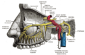
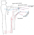
The deep petrosal nerve originates from the sympathetic trunk, specifically from the carotid plexus. It is a postganglionic sympathetic fiber that travels along the internal carotid artery. The nerve then enters the cranial cavity through the carotid canal, and proceeds to join the greater petrosal nerve at the foramen lacerum. This junction forms the nerve of the pterygoid canal (Vidian nerve), which continues to the pterygopalatine ganglion.
Function[edit]
The primary function of the deep petrosal nerve is to provide sympathetic innervation to the lacrimal gland, aiding in tear production. Additionally, it supplies the nasal mucosa, contributing to vasoconstriction and thus playing a role in regulating nasal airflow and mucosal secretion. The nerve also innervates the palate, affecting minor salivary gland secretion.
Clinical Significance[edit]
Understanding the path of the deep petrosal nerve is crucial in various surgical procedures to avoid inadvertent damage. Lesions affecting this nerve can lead to Horner's syndrome, characterized by ptosis (drooping of the upper eyelid), miosis (constricted pupil), and anhidrosis (lack of sweating) on the affected side of the face. Furthermore, its role in tear production makes it a subject of interest in studies related to dry eye syndrome and other lacrimal gland disorders.
Diagnosis and Treatment[edit]
Diagnosis of conditions involving the deep petrosal nerve typically involves a combination of clinical examination and imaging studies, such as MRI or CT scans, to visualize the nerve's pathway and identify any abnormalities. Treatment depends on the underlying cause but may include medications to manage symptoms or surgical intervention in cases where structural anomalies or tumors are present.
See Also[edit]

-
 Deep petrosal nerve
 -
 Deep petrosal nerve
 -
 Deep petrosal nerve


Medical Disclaimer: WikiMD is for informational purposes only and is not a substitute for professional medical advice. Content may be inaccurate or outdated and should not be used for diagnosis or treatment. Always consult your healthcare provider for medical decisions. Verify information with trusted sources such as CDC.gov and NIH.gov. By using this site, you agree that WikiMD is not liable for any outcomes related to its content. See full disclaimer.
Credits:Most images are courtesy of Wikimedia commons, and templates, categories Wikipedia, licensed under CC BY SA or similar.
Translate page: - East Asian
中文,
日本,
한국어,
South Asian
हिन्दी,
தமிழ்,
తెలుగు,
Urdu,
ಕನ್ನಡ,
Southeast Asian
Indonesian,
Vietnamese,
Thai,
မြန်မာဘာသာ,
বাংলা
European
español,
Deutsch,
français,
Greek,
português do Brasil,
polski,
română,
русский,
Nederlands,
norsk,
svenska,
suomi,
Italian
Middle Eastern & African
عربى,
Turkish,
Persian,
Hebrew,
Afrikaans,
isiZulu,
Kiswahili,
Other
Bulgarian,
Hungarian,
Czech,
Swedish,
മലയാളം,
मराठी,
ਪੰਜਾਬੀ,
ગુજરાતી,
Portuguese,
Ukrainian
