Coronary catheterization
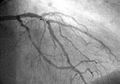
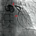
Coronary catheterization is a medical procedure used to diagnose and treat certain cardiovascular conditions. During coronary catheterization, a long thin tube called a catheter is inserted in an artery or vein in your groin, neck or arm and threaded through your blood vessels to your heart.
Procedure[edit]
Coronary catheterization procedures are done in a hospital. You're awake during the procedure, and it may cause little or no pain. The procedure often takes 30 minutes to an hour.
Uses[edit]
Coronary catheterization can help diagnose heart conditions, perform procedures to treat heart disease and check the heart for problems.
Risks[edit]
Coronary catheterization carries a risk of complications, including heart attack, stroke and death. These risks are low.
Preparation[edit]
Before your coronary catheterization, your doctor will give you detailed instructions on how to prepare for your procedure.
Results[edit]
After your coronary catheterization, your doctor will discuss the results with you.
See also[edit]

This WikiMD article can only be edited by registered and verified editors. You can log in or register.
-
 Coronary catheterization
 -
 Coronary angiography
 -
 Coronary stenosis
 -
 Coronary revascularization


Medical Disclaimer: WikiMD is for informational purposes only and is not a substitute for professional medical advice. Content may be inaccurate or outdated and should not be used for diagnosis or treatment. Always consult your healthcare provider for medical decisions. Verify information with trusted sources such as CDC.gov and NIH.gov. By using this site, you agree that WikiMD is not liable for any outcomes related to its content. See full disclaimer.
Credits:Most images are courtesy of Wikimedia commons, and templates, categories Wikipedia, licensed under CC BY SA or similar.
Translate page: - East Asian
中文,
日本,
한국어,
South Asian
हिन्दी,
தமிழ்,
తెలుగు,
Urdu,
ಕನ್ನಡ,
Southeast Asian
Indonesian,
Vietnamese,
Thai,
မြန်မာဘာသာ,
বাংলা
European
español,
Deutsch,
français,
Greek,
português do Brasil,
polski,
română,
русский,
Nederlands,
norsk,
svenska,
suomi,
Italian
Middle Eastern & African
عربى,
Turkish,
Persian,
Hebrew,
Afrikaans,
isiZulu,
Kiswahili,
Other
Bulgarian,
Hungarian,
Czech,
Swedish,
മലയാളം,
मराठी,
ਪੰਜਾਬੀ,
ગુજરાતી,
Portuguese,
Ukrainian
