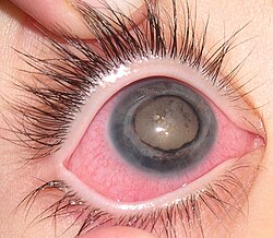
Coats' disease
| Coats' disease | |
|---|---|

| |
| Synonyms | Exudative retinitis, Retinal telangiectasis, Coates' disease |
| Pronounce | N/A |
| Field | Ophthalmology |
| Symptoms | Vision loss, leukocoria, photopsia, floaters |
| Complications | Retinal detachment, glaucoma, cataracts, blindness |
| Onset | Typically in childhood (1st decade of life) |
| Duration | Progressive or self-limiting |
| Types | Unilateral (most common), bilateral (rare) |
| Causes | Abnormal retinal blood vessel development |
| Risks | Male gender, childhood onset |
| Diagnosis | Fundoscopic exam, fluorescein angiography, imaging (US, CT, MRI) |
| Differential diagnosis | Retinoblastoma, Persistent fetal vasculature, Retinal detachment |
| Prevention | None known |
| Treatment | Laser photocoagulation, cryotherapy, vitrectomy |
| Medication | Anti-VEGF therapy (in select cases) |
| Prognosis | Variable; may stabilize, progress, or cause blindness |
| Frequency | 1 in 100,000 |
| Deaths | Rare, if misdiagnosed as retinoblastoma |
Coats' disease is a rare, congenital, and non-hereditary eye disorder that primarily affects young males. It is characterized by abnormal development of retinal blood vessels, leading to fluid leakage, cholesterol deposits, and potential vision loss. In severe cases, it can result in total blindness due to retinal detachment and secondary complications.
Although Coats' disease primarily presents unilaterally (affecting one eye), bilateral cases have been reported. Due to its similarities in presentation to retinoblastoma, careful diagnosis is essential to avoid unnecessary enucleation (eye removal).
Signs and Symptoms[edit]
The symptoms of Coats' disease vary depending on the stage of progression. The earliest symptom is often leukocoria (an abnormal white reflection from the retina), which is frequently first noticed in flash photography.
Common symptoms include:
- Leukocoria – White or yellow pupil reflection in photographs.
- Blurred vision – Often noticed when covering the unaffected eye.
- Loss of depth perception – Due to one-eye compensation.
- Photopsia – Flashing lights in the affected eye.
- Floaters – Small moving spots in vision.
- Peripheral vision loss – Often starts in the upper field due to fluid pooling in the lower retina.
- Painless progression – Until complications such as glaucoma cause discomfort.
Coats' disease is often detected incidentally when parents notice a child with yellow-eye reflex in photos rather than the usual red-eye effect. Early evaluation by an ophthalmologist is critical for diagnosis.

Advanced Symptoms[edit]
If left untreated, the disease may progress to:
- Exudative retinal detachment – Separation of the retina from its underlying support.
- Glaucoma – Increased eye pressure due to fluid buildup.
- Cataracts – Clouding of the lens.
- Painful eye swelling – If fluid drainage is obstructed.
- Blindness – If retinal detachment or optic nerve damage occurs.
Causes and Risk Factors[edit]
The exact cause of Coats' disease is unknown, but it is believed to result from defective blood vessel formation in the retina. The disease occurs sporadically and is not inherited.
Risk Factors[edit]
- Male sex – About 90% of cases occur in males.
- Childhood onset – Most common between ages 6 and 8.
- Unilateral occurrence – Affects one eye in most cases.
Pathogenesis[edit]
Coats' disease arises due to leakage from defective capillaries in the retina. This leads to:
- Cholesterol and lipid deposits within retinal layers.
- Thickening of the retina due to protein buildup.
- Gradual detachment of the retina as fluid accumulates.
As the disease progresses, these changes disrupt normal vision and may eventually lead to permanent vision loss if untreated.
Diagnosis[edit]
== Clinical Examination Diagnosis is confirmed through fundoscopic examination by an ophthalmologist. Key findings include:
- Tortuous and dilated blood vessels in the retina.
- Retinal exudates (lipid deposits).
- Varying degrees of retinal detachment.
Imaging Studies[edit]

Additional tests help distinguish Coats' disease from retinoblastoma and other retinal disorders:
- Fluorescein Angiography – Evaluates leaking retinal blood vessels.
- Ultrasound (US) – Detects fluid accumulation and detachment.
- Computed Tomography (CT) – Shows a hyperdense vitreous.
- Magnetic Resonance Imaging (MRI) – Identifies fluid leakage and retinal thickening.
Treatment[edit]
Early-Stage Treatment[edit]
When diagnosed early, treatment can halt disease progression and preserve vision. Common approaches include:
- Laser photocoagulation – Seals leaking blood vessels.
- Cryotherapy – Freezes and destroys abnormal vessels.
- Anti-VEGF injections – May help reduce fluid accumulation.
Advanced Disease Management[edit]
For severe cases with retinal detachment, more invasive treatments are required:
- Vitrectomy – Surgical removal of vitreous fluid to reattach the retina.
- Scleral Buckle – A silicone band placed around the eye to support the retina.
- Enucleation (Eye Removal) – Performed only if the eye is non-functional and painful.
Prognosis[edit]
The prognosis of Coats' disease varies based on stage at diagnosis and treatment response.
- Mild Cases – If caught early, vision can be preserved or stabilized.
- Moderate Cases – May result in permanent vision impairment.
- Severe Cases – Often lead to blindness or eye loss.
Some cases stabilize on their own, while others progress rapidly, requiring aggressive intervention.
Historical Background[edit]
Coats' disease is named after George Coats, who first described the condition in 1908.
See Also[edit]
External Links[edit]
Ad. Transform your life with W8MD's Budget GLP-1 injections from $75


W8MD offers a medical weight loss program to lose weight in Philadelphia. Our physician-supervised medical weight loss provides:
- Weight loss injections in NYC (generic and brand names):
- Zepbound / Mounjaro, Wegovy / Ozempic, Saxenda
- Most insurances accepted or discounted self-pay rates. We will obtain insurance prior authorizations if needed.
- Generic GLP1 weight loss injections from $75 for the starting dose.
- Also offer prescription weight loss medications including Phentermine, Qsymia, Diethylpropion, Contrave etc.
NYC weight loss doctor appointmentsNYC weight loss doctor appointments
Start your NYC weight loss journey today at our NYC medical weight loss and Philadelphia medical weight loss clinics.
- Call 718-946-5500 to lose weight in NYC or for medical weight loss in Philadelphia 215-676-2334.
- Tags:NYC medical weight loss, Philadelphia lose weight Zepbound NYC, Budget GLP1 weight loss injections, Wegovy Philadelphia, Wegovy NYC, Philadelphia medical weight loss, Brookly weight loss and Wegovy NYC
|
WikiMD's Wellness Encyclopedia |
| Let Food Be Thy Medicine Medicine Thy Food - Hippocrates |
Medical Disclaimer: WikiMD is not a substitute for professional medical advice. The information on WikiMD is provided as an information resource only, may be incorrect, outdated or misleading, and is not to be used or relied on for any diagnostic or treatment purposes. Please consult your health care provider before making any healthcare decisions or for guidance about a specific medical condition. WikiMD expressly disclaims responsibility, and shall have no liability, for any damages, loss, injury, or liability whatsoever suffered as a result of your reliance on the information contained in this site. By visiting this site you agree to the foregoing terms and conditions, which may from time to time be changed or supplemented by WikiMD. If you do not agree to the foregoing terms and conditions, you should not enter or use this site. See full disclaimer.
Credits:Most images are courtesy of Wikimedia commons, and templates, categories Wikipedia, licensed under CC BY SA or similar.
Translate this page: - East Asian
中文,
日本,
한국어,
South Asian
हिन्दी,
தமிழ்,
తెలుగు,
Urdu,
ಕನ್ನಡ,
Southeast Asian
Indonesian,
Vietnamese,
Thai,
မြန်မာဘာသာ,
বাংলা
European
español,
Deutsch,
français,
Greek,
português do Brasil,
polski,
română,
русский,
Nederlands,
norsk,
svenska,
suomi,
Italian
Middle Eastern & African
عربى,
Turkish,
Persian,
Hebrew,
Afrikaans,
isiZulu,
Kiswahili,
Other
Bulgarian,
Hungarian,
Czech,
Swedish,
മലയാളം,
मराठी,
ਪੰਜਾਬੀ,
ગુજરાતી,
Portuguese,
Ukrainian


