Aortic arches
-
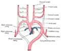 Diagram of the aortic arches in a human embryo.
 -
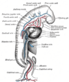 Illustration showing the development of the aortic arches.
 -
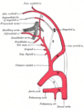 Aortic arches in a human embryo, lateral view.


Aortic Arches[edit]
The aortic arches are a series of six paired embryological vascular structures that give rise to several major arteries in the adult. These arches are crucial in the development of the circulatory system in vertebrates, including humans. They are also known as pharyngeal arch arteries because they are associated with the pharyngeal arches.
Development[edit]
The aortic arches develop from the aortic sac, which is an extension of the truncus arteriosus. During embryogenesis, these arches form between the ventral and dorsal aortae and are initially symmetrical. However, as development progresses, they undergo significant remodeling to form the asymmetric arterial system seen in adults.
First Aortic Arch[edit]
The first aortic arch largely regresses, but it contributes to the formation of the maxillary artery, which supplies the deep structures of the face.
Second Aortic Arch[edit]
The second aortic arch also regresses significantly, with remnants forming the stapedial artery in the embryo, which typically regresses in humans.
Third Aortic Arch[edit]
The third aortic arch is crucial as it forms the common carotid arteries and the proximal portion of the internal carotid arteries.
Fourth Aortic Arch[edit]
The fourth aortic arch has different fates on the left and right sides. On the left, it forms part of the aortic arch itself, while on the right, it contributes to the formation of the right subclavian artery.
Fifth Aortic Arch[edit]
The fifth aortic arch is often absent or rudimentary in humans and does not contribute significantly to the adult arterial system.
Sixth Aortic Arch[edit]
The sixth aortic arch is also known as the pulmonary arch. It gives rise to the pulmonary arteries and the ductus arteriosus, which connects the pulmonary trunk to the aortic arch in the fetus. The ductus arteriosus typically closes after birth to become the ligamentum arteriosum.
Clinical Significance[edit]
Abnormal development of the aortic arches can lead to congenital vascular anomalies. These include conditions such as coarctation of the aorta, interrupted aortic arch, and various forms of vascular rings that can compress the trachea or esophagus.
Related Pages[edit]
| Human embryogenesis in the first three weeks | ||||||
|---|---|---|---|---|---|---|
|
| Arteries and veins | ||||||||||||
|---|---|---|---|---|---|---|---|---|---|---|---|---|
|

Medical Disclaimer: WikiMD is for informational purposes only and is not a substitute for professional medical advice. Content may be inaccurate or outdated and should not be used for diagnosis or treatment. Always consult your healthcare provider for medical decisions. Verify information with trusted sources such as CDC.gov and NIH.gov. By using this site, you agree that WikiMD is not liable for any outcomes related to its content. See full disclaimer.
Credits:Most images are courtesy of Wikimedia commons, and templates, categories Wikipedia, licensed under CC BY SA or similar.
Translate page: - East Asian
中文,
日本,
한국어,
South Asian
हिन्दी,
தமிழ்,
తెలుగు,
Urdu,
ಕನ್ನಡ,
Southeast Asian
Indonesian,
Vietnamese,
Thai,
မြန်မာဘာသာ,
বাংলা
European
español,
Deutsch,
français,
Greek,
português do Brasil,
polski,
română,
русский,
Nederlands,
norsk,
svenska,
suomi,
Italian
Middle Eastern & African
عربى,
Turkish,
Persian,
Hebrew,
Afrikaans,
isiZulu,
Kiswahili,
Other
Bulgarian,
Hungarian,
Czech,
Swedish,
മലയാളം,
मराठी,
ਪੰਜਾਬੀ,
ગુજરાતી,
Portuguese,
Ukrainian
