Air bronchogram
Radiological sign of air-filled bronchi on a background of opaque lung tissue
An air bronchogram is a radiological sign seen on a chest X-ray or CT scan of the lungs. It is characterized by the presence of air-filled bronchi that are made visible by the opacification of surrounding alveoli. This sign is typically indicative of lung consolidation, which can occur in various conditions such as pneumonia, pulmonary edema, or atelectasis.
Pathophysiology[edit]
The air bronchogram sign occurs when the alveoli, which are normally air-filled, become filled with fluid, pus, blood, or cells, leading to increased lung opacity. The bronchi, which remain air-filled, stand out against the opaque background. This contrast allows the bronchi to be visualized as dark branching structures on imaging studies.
Clinical significance[edit]
Air bronchograms are most commonly associated with conditions that cause lung consolidation. Some of the common causes include:
- Pneumonia: Infections of the lung parenchyma can lead to consolidation and the presence of air bronchograms.
- Pulmonary edema: Fluid accumulation in the alveoli due to heart failure or other causes can result in this sign.
- Atelectasis: Collapse of lung tissue can also present with air bronchograms, although the pattern may vary.
- Acute respiratory distress syndrome (ARDS): This severe inflammatory condition can lead to widespread lung consolidation.
Imaging techniques[edit]
Air bronchograms are best visualized using:
- Chest X-ray: A standard imaging technique that can reveal air bronchograms in cases of lung consolidation.
- Computed tomography (CT) scan: Provides a more detailed view of the lung structures and is more sensitive in detecting air bronchograms.
Interpretation[edit]
The presence of air bronchograms on imaging studies suggests that the bronchi are patent and that the surrounding alveoli are filled with material other than air. This finding helps differentiate between different types of lung pathology and can guide further diagnostic and therapeutic interventions.
Related pages[edit]
Gallery[edit]
-
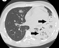 CT scan showing air bronchograms in Legionnaires' disease
 -
 Creative Commons icon


Medical Disclaimer: WikiMD is for informational purposes only and is not a substitute for professional medical advice. Content may be inaccurate or outdated and should not be used for diagnosis or treatment. Always consult your healthcare provider for medical decisions. Verify information with trusted sources such as CDC.gov and NIH.gov. By using this site, you agree that WikiMD is not liable for any outcomes related to its content. See full disclaimer.
Credits:Most images are courtesy of Wikimedia commons, and templates, categories Wikipedia, licensed under CC BY SA or similar.
Translate page: - East Asian
中文,
日本,
한국어,
South Asian
हिन्दी,
தமிழ்,
తెలుగు,
Urdu,
ಕನ್ನಡ,
Southeast Asian
Indonesian,
Vietnamese,
Thai,
မြန်မာဘာသာ,
বাংলা
European
español,
Deutsch,
français,
Greek,
português do Brasil,
polski,
română,
русский,
Nederlands,
norsk,
svenska,
suomi,
Italian
Middle Eastern & African
عربى,
Turkish,
Persian,
Hebrew,
Afrikaans,
isiZulu,
Kiswahili,
Other
Bulgarian,
Hungarian,
Czech,
Swedish,
മലയാളം,
मराठी,
ਪੰਜਾਬੀ,
ગુજરાતી,
Portuguese,
Ukrainian
