Acoustic angiography
Acoustic Angiography

Acoustic angiography is an advanced ultrasound imaging technique used to visualize the vascular system with high resolution and specificity. This method leverages the unique properties of microbubbles as contrast agents to enhance the imaging of blood vessels.
Principles[edit]
Acoustic angiography utilizes the nonlinear response of microbubbles to ultrasound waves. When exposed to ultrasound, these microbubbles oscillate and produce harmonic signals that can be detected and used to create detailed images of the vasculature. This technique is particularly useful for imaging small vessels and detecting abnormalities in the vascular structure.
Technology[edit]
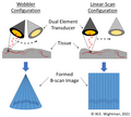
The key component of acoustic angiography is the dual-element transducer, which consists of separate elements for transmitting and receiving ultrasound signals. This configuration allows for the isolation of harmonic signals generated by the microbubbles, enhancing the contrast and resolution of the images.
Scanning Configurations[edit]
There are different scanning configurations used in acoustic angiography, including wobbler and linear scanning.
- Wobbler Scanning: In this configuration, the transducer is mechanically wobbled to cover a larger area. This method is beneficial for imaging larger regions but may have limitations in terms of speed and resolution.
- Linear Scanning: This configuration involves moving the transducer linearly across the area of interest. It provides high-resolution images and is suitable for detailed studies of specific vascular regions.
Applications[edit]
Acoustic angiography is used in various medical fields, including:
- Cardiology: For imaging coronary arteries and detecting blockages or abnormalities.
- Oncology: To visualize tumor vasculature and assess the effectiveness of anti-angiogenic therapies.
- Neurology: For imaging cerebral vessels and diagnosing conditions such as aneurysms or arteriovenous malformations.
Advantages[edit]
The main advantages of acoustic angiography include:
- High spatial resolution, allowing for detailed visualization of small vessels.
- Non-invasive nature, reducing the risk associated with traditional angiography methods.
- Enhanced contrast due to the use of microbubbles, improving the detection of vascular abnormalities.
Limitations[edit]
Despite its advantages, acoustic angiography has some limitations:
- Limited penetration depth, which may restrict its use in imaging deep-seated vessels.
- Dependence on the availability and stability of microbubble contrast agents.
Future Directions[edit]
Research is ongoing to improve the technology and expand its clinical applications. Innovations in transducer design and microbubble formulation are expected to enhance the capabilities of acoustic angiography.
Related Pages[edit]
-
Acoustic_angiography
-
Acoustic_angiography
Ad. Transform your life with W8MD's Budget GLP-1 injections from $75


W8MD offers a medical weight loss program to lose weight in Philadelphia. Our physician-supervised medical weight loss provides:
- Weight loss injections in NYC (generic and brand names):
- Zepbound / Mounjaro, Wegovy / Ozempic, Saxenda
- Most insurances accepted or discounted self-pay rates. We will obtain insurance prior authorizations if needed.
- Generic GLP1 weight loss injections from $75 for the starting dose.
- Also offer prescription weight loss medications including Phentermine, Qsymia, Diethylpropion, Contrave etc.
NYC weight loss doctor appointmentsNYC weight loss doctor appointments
Start your NYC weight loss journey today at our NYC medical weight loss and Philadelphia medical weight loss clinics.
- Call 718-946-5500 to lose weight in NYC or for medical weight loss in Philadelphia 215-676-2334.
- Tags:NYC medical weight loss, Philadelphia lose weight Zepbound NYC, Budget GLP1 weight loss injections, Wegovy Philadelphia, Wegovy NYC, Philadelphia medical weight loss, Brookly weight loss and Wegovy NYC
|
WikiMD's Wellness Encyclopedia |
| Let Food Be Thy Medicine Medicine Thy Food - Hippocrates |
Medical Disclaimer: WikiMD is not a substitute for professional medical advice. The information on WikiMD is provided as an information resource only, may be incorrect, outdated or misleading, and is not to be used or relied on for any diagnostic or treatment purposes. Please consult your health care provider before making any healthcare decisions or for guidance about a specific medical condition. WikiMD expressly disclaims responsibility, and shall have no liability, for any damages, loss, injury, or liability whatsoever suffered as a result of your reliance on the information contained in this site. By visiting this site you agree to the foregoing terms and conditions, which may from time to time be changed or supplemented by WikiMD. If you do not agree to the foregoing terms and conditions, you should not enter or use this site. See full disclaimer.
Credits:Most images are courtesy of Wikimedia commons, and templates, categories Wikipedia, licensed under CC BY SA or similar.
Translate this page: - East Asian
中文,
日本,
한국어,
South Asian
हिन्दी,
தமிழ்,
తెలుగు,
Urdu,
ಕನ್ನಡ,
Southeast Asian
Indonesian,
Vietnamese,
Thai,
မြန်မာဘာသာ,
বাংলা
European
español,
Deutsch,
français,
Greek,
português do Brasil,
polski,
română,
русский,
Nederlands,
norsk,
svenska,
suomi,
Italian
Middle Eastern & African
عربى,
Turkish,
Persian,
Hebrew,
Afrikaans,
isiZulu,
Kiswahili,
Other
Bulgarian,
Hungarian,
Czech,
Swedish,
മലയാളം,
मराठी,
ਪੰਜਾਬੀ,
ગુજરાતી,
Portuguese,
Ukrainian

