Nasal glial heterotopia
| Nasal glial heterotopia | |
|---|---|
| Synonyms | Nasal glioma |
| Pronounce | N/A |
| Specialty | N/A |
| Symptoms | Nasal obstruction, mass in the nasal region |
| Complications | Infection, respiratory distress |
| Onset | Congenital |
| Duration | Lifelong unless surgically removed |
| Types | N/A |
| Causes | Developmental anomaly |
| Risks | |
| Diagnosis | MRI, CT scan, Biopsy |
| Differential diagnosis | N/A |
| Prevention | N/A |
| Treatment | Surgical excision |
| Medication | N/A |
| Prognosis | Excellent with treatment |
| Frequency | Rare |
| Deaths | N/A |
Nasal glial heterotopia, also known as nasal glioma, is a rare congenital condition characterized by the presence of glial tissue in the nasal region. This condition is a type of heterotopia, where normal tissue is found in an abnormal location.
Pathophysiology[edit]
Nasal glial heterotopia arises due to a developmental anomaly during embryogenesis. During the formation of the central nervous system, glial tissue may become sequestered outside the cranial cavity. This ectopic glial tissue can then become embedded in the nasal region, leading to the formation of a mass. Unlike true neoplasms, nasal glial heterotopia does not exhibit proliferative growth.
Clinical Presentation[edit]
Patients with nasal glial heterotopia typically present with a mass in the nasal region. Common symptoms include:
- Nasal obstruction
- Visible mass or swelling on the nose or nasal bridge
- Respiratory distress in severe cases
The mass is usually firm, non-compressible, and non-pulsatile. It is important to differentiate nasal glial heterotopia from other nasal masses such as encephalocele or dermoid cyst.
Diagnosis[edit]
The diagnosis of nasal glial heterotopia involves a combination of imaging studies and histological examination. Key diagnostic tools include:
- Magnetic Resonance Imaging (MRI): Provides detailed images of the nasal mass and its relation to surrounding structures.
- Computed Tomography (CT) scan: Helps in assessing any bony involvement or defects.
- Biopsy: Confirms the presence of glial tissue histologically.
Treatment[edit]
The primary treatment for nasal glial heterotopia is surgical excision. The goal of surgery is to completely remove the ectopic glial tissue while preserving normal nasal structures. Surgical approaches may vary depending on the size and location of the mass.
Prognosis[edit]
The prognosis for patients with nasal glial heterotopia is excellent following complete surgical excision. Recurrence is rare, and most patients experience resolution of symptoms post-surgery.
Epidemiology[edit]
Nasal glial heterotopia is a rare condition, with only a few cases reported in the medical literature. It is typically diagnosed in infancy or early childhood, although cases in adults have been documented.
See also[edit]
External links[edit]
- [Link to relevant medical resources]
Template:Congenital malformations of the nervous system
-
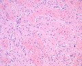 Nasal glial heterotopia hematoxylin and eosin stained photomicrograph
 -
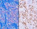 Nasal glial heterotopia stained with trichrome and GFAP
 -
 Nasal glial heterotopia computed tomography scan

Ad. Transform your health with W8MD Weight Loss, Sleep & MedSpa

Tired of being overweight?
Special offer:
Budget GLP-1 weight loss medications
- Semaglutide starting from $29.99/week and up with insurance for visit of $59.99 and up per week self pay.
- Tirzepatide starting from $45.00/week and up (dose dependent) or $69.99/week and up self pay
✔ Same-week appointments, evenings & weekends
Learn more:
- GLP-1 weight loss clinic NYC
- W8MD's NYC medical weight loss
- W8MD Philadelphia GLP-1 shots
- Philadelphia GLP-1 injections
- Affordable GLP-1 shots NYC
|
WikiMD Medical Encyclopedia |
Medical Disclaimer: WikiMD is for informational purposes only and is not a substitute for professional medical advice. Content may be inaccurate or outdated and should not be used for diagnosis or treatment. Always consult your healthcare provider for medical decisions. Verify information with trusted sources such as CDC.gov and NIH.gov. By using this site, you agree that WikiMD is not liable for any outcomes related to its content. See full disclaimer.
Credits:Most images are courtesy of Wikimedia commons, and templates, categories Wikipedia, licensed under CC BY SA or similar.
Translate this page: - East Asian
中文,
日本,
한국어,
South Asian
हिन्दी,
தமிழ்,
తెలుగు,
Urdu,
ಕನ್ನಡ,
Southeast Asian
Indonesian,
Vietnamese,
Thai,
မြန်မာဘာသာ,
বাংলা
European
español,
Deutsch,
français,
Greek,
português do Brasil,
polski,
română,
русский,
Nederlands,
norsk,
svenska,
suomi,
Italian
Middle Eastern & African
عربى,
Turkish,
Persian,
Hebrew,
Afrikaans,
isiZulu,
Kiswahili,
Other
Bulgarian,
Hungarian,
Czech,
Swedish,
മലയാളം,
मराठी,
ਪੰਜਾਬੀ,
ગુજરાતી,
Portuguese,
Ukrainian
