Urachal cancer: Difference between revisions
CSV import |
CSV import |
||
| Line 31: | Line 31: | ||
[[Category:Rare cancers]] | [[Category:Rare cancers]] | ||
[[Category:Oncology]] | [[Category:Oncology]] | ||
<gallery> | |||
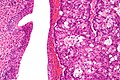
File:Urachal carcinoma - high mag.jpg|Urachal cancer | |||
File:UrC CDX2 200x.tif|Urachal cancer | |||
</gallery> | |||
Revision as of 01:36, 20 February 2025
A rare type of cancer originating from the urachus
Urachal cancer is a rare form of cancer that arises from the urachus, a vestigial remnant of the allantois that connects the bladder to the umbilicus during fetal development. This type of cancer is most commonly found in adults and is often diagnosed at an advanced stage due to its asymptomatic nature in the early stages.
Anatomy and Physiology
The urachus is a fibrous cord that is a remnant of the allantois, which is part of the fetal urinary tract. During fetal development, the urachus connects the bladder to the umbilical cord, allowing urine to drain from the bladder into the amniotic sac. After birth, the urachus typically obliterates and becomes the median umbilical ligament. However, in some individuals, remnants of the urachus persist, which can lead to the development of urachal abnormalities, including urachal cancer.
Pathophysiology
Urachal cancer is believed to arise from metaplasia of the epithelial cells lining the urachus. The most common histological type of urachal cancer is adenocarcinoma, which accounts for approximately 90% of cases. Other types include squamous cell carcinoma and transitional cell carcinoma. The cancer typically presents as a mass at the dome of the bladder and can invade surrounding structures.
Clinical Presentation
Patients with urachal cancer may present with nonspecific symptoms such as hematuria (blood in the urine), abdominal pain, or a palpable mass. Due to the deep location of the urachus, symptoms often do not appear until the cancer is advanced. Some patients may also experience urinary tract infections or mucusuria (mucus in the urine).
Diagnosis
The diagnosis of urachal cancer involves a combination of imaging studies and histological examination. Ultrasound, CT scan, and MRI are commonly used to visualize the tumor and assess its extent. A cystoscopy may be performed to evaluate the bladder and obtain a biopsy of the tumor. Histological examination of the biopsy is necessary to confirm the diagnosis and determine the type of cancer.
Treatment
The primary treatment for urachal cancer is surgical resection. A procedure known as a partial cystectomy is often performed, which involves removing the tumor along with a portion of the bladder. In some cases, a radical cystectomy may be necessary. Chemotherapy and radiation therapy may be used as adjunctive treatments, particularly in cases where the cancer has metastasized or is not amenable to complete surgical resection.
Prognosis
The prognosis for urachal cancer depends on the stage at diagnosis and the completeness of surgical resection. Early-stage cancers that are completely resected have a better prognosis, while advanced-stage cancers with metastasis have a poorer outcome. Regular follow-up is essential to monitor for recurrence.
Related pages
-
 Urachal cancer
 -
 Urachal cancer

