Ventricular system: Difference between revisions
CSV import |
CSV import |
||
| Line 36: | Line 36: | ||
[[Category:Neuroanatomy]] | [[Category:Neuroanatomy]] | ||
[[Category:Ventricular system]] | [[Category:Ventricular system]] | ||
<gallery> | |||
File:Brain-ventricle-anatomy-diagram.jpg|Diagram of brain ventricles | |||
File:Human_ventricular_system_-_animation.gif|Animation of human ventricular system | |||
File:Blausen_0896_Ventricles_Brain.png|Illustration of brain ventricles | |||
File:NPH_MRI_272_GILD.gif|MRI of normal pressure hydrocephalus | |||
File:Ventricular_system.stl|3D model of the ventricular system | |||
File:Gray734.png|Gray's anatomy illustration of ventricles | |||
File:Lateral_Ventricles_-_DK_ATLAS.png|Atlas image of lateral ventricles | |||
</gallery> | |||
Latest revision as of 11:21, 18 February 2025
A set of structures in the brain responsible for the production and circulation of cerebrospinal fluid
Ventricular system[edit]
The ventricular system is a set of four interconnected cavities (ventricles) in the brain, where cerebrospinal fluid (CSF) is produced and circulated. The system is composed of the two lateral ventricles, the third ventricle, and the fourth ventricle. These structures are crucial for protecting the brain, providing buoyancy, and removing waste products.

Anatomy[edit]
The ventricular system is lined with ependymal cells, which are a type of glial cell that helps in the production and circulation of CSF. The lateral ventricles are the largest and are located in the cerebral hemispheres. They connect to the third ventricle via the interventricular foramina (foramina of Monro). The third ventricle is situated in the midline of the brain, between the two halves of the thalamus. It connects to the fourth ventricle via the cerebral aqueduct (aqueduct of Sylvius).
The fourth ventricle is located between the pons and the cerebellum. It is continuous with the central canal of the spinal cord and opens into the subarachnoid space through the median aperture (foramen of Magendie) and the two lateral apertures (foramina of Luschka).

Function[edit]
The primary function of the ventricular system is the production and circulation of CSF, which cushions the brain and spinal cord, maintains intracranial pressure, and removes metabolic waste. CSF is produced by the choroid plexus, a network of capillaries located in the ventricles.
Clinical significance[edit]
Disorders of the ventricular system can lead to conditions such as hydrocephalus, where there is an abnormal accumulation of CSF, leading to increased intracranial pressure. This can result from blockages in the ventricular system, overproduction of CSF, or impaired absorption. Normal pressure hydrocephalus is a specific type of hydrocephalus that occurs in older adults and is characterized by gait disturbance, dementia, and urinary incontinence.

Related pages[edit]
References[edit]
- Susan,
 Gray's Anatomy: The Anatomical Basis of Clinical Practice,
 41st edition,
 Elsevier,
 2016,


 ISBN 978-0-7020-5230-9,
- ,
 The pathophysiology of idiopathic normal pressure hydrocephalus: Cerebral hydrodynamics and the role of the transmantle pressure gradient,
 Fluids and Barriers of the CNS,
 2000,
 Vol. 7(Issue: 1),
 pp. 1-11,
 DOI: 10.1186/2045-8118-7-1,
-
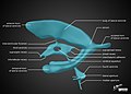 Diagram of brain ventricles
 -
 Animation of human ventricular system
 -
 Illustration of brain ventricles
 -
 MRI of normal pressure hydrocephalus
 -
 3D model of the ventricular system
 -
 Gray's anatomy illustration of ventricles
 -
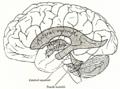
 Atlas image of lateral ventricles


