Podocyte: Difference between revisions
CSV import Tags: mobile edit mobile web edit |
CSV import |
||
| Line 41: | Line 41: | ||
{{Kidney}} | {{Kidney}} | ||
{{Cell-biology-stub}} | {{Cell-biology-stub}} | ||
<gallery> | |||
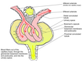
File:Renal_corpuscle-en.svg|Renal corpuscle diagram | |||
File:Glomerular_Physiology.png|Glomerular physiology | |||
File:Glomerulum_of_mouse_kidney_in_Scanning_Electron_Microscope,_magnification_5,000x.GIF|Glomerulum of mouse kidney in Scanning Electron Microscope, magnification 5,000x | |||
File:Filtration_barrier.svg|Filtration barrier | |||
File:Podo001.jpg|Podocyte | |||
File:Morphologic_patterns_of_podocyte_injury.jpg|Morphologic patterns of podocyte injury | |||
</gallery> | |||
Latest revision as of 11:02, 18 February 2025
Podocyte
A Podocyte is a type of cell found in the kidney, specifically in the Bowman's capsule of the glomerulus. These cells play a crucial role in the body's ability to filter blood and create urine.
Structure[edit]
Podocytes are unique in their structure, featuring a large cell body with primary processes and secondary foot processes, or pedicels. These foot processes interdigitate with those of neighboring podocytes and are connected by a thin filtration slit diaphragm. This structure allows for the efficient filtration of blood.
Function[edit]
The primary function of podocytes is to filter blood in the glomerulus, a network of small blood vessels in the kidney. They do this by forming a barrier that prevents the passage of large molecules, such as proteins, while allowing smaller molecules, such as water and salts, to pass through. This filtration process is essential for the production of urine.
Clinical significance[edit]
Damage to podocytes can lead to a number of kidney diseases, including Focal segmental glomerulosclerosis (FSGS) and Minimal change disease (MCD). These conditions can result in proteinuria, the presence of excess proteins in the urine, and can ultimately lead to kidney failure.
Research[edit]
Research into podocytes has increased our understanding of kidney function and disease. For example, studies have shown that podocytes can regenerate and repair themselves, which has implications for the treatment of kidney diseases. Furthermore, research is ongoing into the genetic and molecular mechanisms that regulate podocyte function and health.
See also[edit]
This medical article is a stub. You can help WikiMD by expanding the page. |
References[edit]
<references />
External links[edit]
- Podocyte Biology and Pathogenesis of Kidney Disease at the US National Library of Medicine
- Podocyte-specific knockout of the adaptor protein CD2-associated protein promotes proteinuria and glomerulosclerosis in mice at Kidney International
| Urinary system - Kidney - edit |
|---|
| Renal capsule | Renal cortex | Renal medulla (Renal sinus, Renal pyramids) | Renal calyx | Renal pelvis |
| Nephron - Renal corpuscle (Glomerulus, Bowman's capsule) → Proximal tubule → Loop of Henle → Distal convoluted tubule → Collecting ducts
Juxtaglomerular apparatus (Macula densa, Juxtaglomerular cells) Renal circulation - Renal artery → Interlobar arteries → Arcuate arteries → Cortical radial arteries → Afferent arterioles → Glomerulus → Efferent arterioles → Vasa recta → Arcuate vein → Renal vein |
| Renal physiology |
| Filtration - Ultrafiltration | Countercurrent exchange
Hormones effecting filtration - Antidiuretic hormone (ADH) | Aldosterone | Atrial natriuretic peptide Endocrine - Renin | Erythropoietin (EPO) | Calcitriol (Active vitamin D) | Prostaglandins |
| Assessing Renal function / Measures of Dialysis |
| Glomerular filtration rate | Creatinine clearance | Renal clearance ratio | Urea reduction ratio | Kt/V | Standardized Kt/V | Hemodialysis product |
This cell biology article is a stub. You can help WikiMD by expanding the page. |
-
 Renal corpuscle diagram
 -
 Glomerular physiology
 -
 Glomerulum of mouse kidney in Scanning Electron Microscope, magnification 5,000x
 -
 Filtration barrier
 -
 Podocyte
 -
 Morphologic patterns of podocyte injury


