Central retinal vein occlusion: Difference between revisions
CSV import |
CSV import |
||
| Line 44: | Line 44: | ||
[[Category:Medical conditions related to obesity]] | [[Category:Medical conditions related to obesity]] | ||
{{medicine-stub}} | {{medicine-stub}} | ||
<gallery> | |||
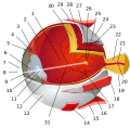
File:Eye-diagram_no_circles_border_1.svg|Central retinal vein occlusion | |||
</gallery> | |||
Revision as of 01:57, 18 February 2025
Central Retinal Vein Occlusion (CRVO) is a condition characterized by the blockage of the central vein in the retina, which is responsible for draining blood from the retina back to the heart. This blockage can lead to a buildup of blood and fluid in the retina, causing vision loss or blindness if not treated promptly. CRVO is one of the most common retinal vascular disorders and is a significant cause of vision loss worldwide.
Causes and Risk Factors
The primary cause of CRVO is the thrombosis (formation of a blood clot) in the central retinal vein, which can be precipitated by various systemic conditions. Key risk factors include:
- Hypertension: High blood pressure can damage the walls of the retinal veins, making them more susceptible to blockage.
- Diabetes mellitus: Diabetes can lead to changes in the blood vessels, including those in the eye, increasing the risk of CRVO.
- Glaucoma: Increased eye pressure can compress the central retinal vein, leading to occlusion.
- Hypercoagulability: Conditions that make the blood more likely to clot can predispose individuals to CRVO.
- Age: The risk of CRVO increases with age, particularly in individuals over the age of 50.
- Smoking: Tobacco use can affect blood flow and contribute to the development of blood clots.
Symptoms
Symptoms of CRVO can vary depending on the severity of the blockage but may include:
- Sudden, painless vision loss in one eye
- Blurred or distorted vision
- Dark spots or lines (floaters) in the vision
- A noticeable difference in color perception between the two eyes
Diagnosis
Diagnosis of CRVO involves a comprehensive eye examination, including:
- Visual acuity test: To assess the extent of vision loss.
- Fundus photography: To capture detailed images of the retina and identify any abnormalities.
- Fluorescein angiography: A test that uses a special dye to highlight the blood vessels in the retina, allowing for the identification of any blockages or leakages.
- Optical coherence tomography (OCT): This non-invasive imaging test provides cross-sectional images of the retina, helping to evaluate the extent of swelling or fluid buildup.
Treatment
Treatment for CRVO aims to manage the underlying condition and prevent further vision loss. Options may include:
- Intravitreal injections: Medications such as anti-VEGF agents or corticosteroids can be injected into the eye to reduce swelling and improve blood flow.
- Laser therapy: Laser treatment can help seal leaking blood vessels and reduce swelling in the retina.
- Management of underlying systemic conditions: Controlling blood pressure, blood sugar levels, and other related conditions is crucial in preventing further damage.
Prognosis
The prognosis for CRVO varies widely and depends on the severity of the occlusion and the promptness of treatment. While some individuals may experience significant improvement in vision, others may suffer from permanent vision loss. Early detection and management of risk factors are key to preventing CRVO and preserving vision.
Prevention
Preventive measures for CRVO primarily involve controlling the risk factors associated with its development. Regular eye examinations, managing systemic conditions like hypertension and diabetes, and adopting a healthy lifestyle can significantly reduce the risk of CRVO.

-
 Central retinal vein occlusion

