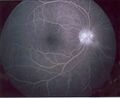
Fluorescein angiography
Fluorescein angiography is a medical procedure used to visualize the blood vessels in the retina and choroid layers of the eye. This procedure involves the injection of a fluorescent dye, called fluorescein, into the bloodstream. The dye travels to the blood vessels in the eye, allowing them to be photographed.
Procedure[edit]
The procedure begins with the administration of a local anesthetic to numb the eye. The fluorescein dye is then injected into a vein in the arm. The dye travels through the bloodstream to the blood vessels in the eye. A special camera equipped with filters that highlight the dye is used to take photographs of the blood vessels as the dye passes through them. The images can then be analyzed to identify any abnormalities in the blood vessels.
Uses[edit]
Fluorescein angiography is used to diagnose and monitor a number of eye conditions, including diabetic retinopathy, macular degeneration, and retinal vein occlusion. It can also be used to evaluate the effectiveness of treatments for these conditions.
Risks[edit]
While fluorescein angiography is generally safe, there are some risks associated with the procedure. These include allergic reactions to the dye, which can cause itching, hives, or in rare cases, anaphylaxis. Other potential complications include nausea, vomiting, and temporary discoloration of the skin and urine.
See also[edit]

This WikiMD article can only be edited by registered and verified editors. You can log in or register.
-
 Fluorescein angiography


Medical Disclaimer: WikiMD is for informational purposes only and is not a substitute for professional medical advice. Content may be inaccurate or outdated and should not be used for diagnosis or treatment. Always consult your healthcare provider for medical decisions. Verify information with trusted sources such as CDC.gov and NIH.gov. By using this site, you agree that WikiMD is not liable for any outcomes related to its content. See full disclaimer.
Credits:Most images are courtesy of Wikimedia commons, and templates, categories Wikipedia, licensed under CC BY SA or similar.
Translate page: - East Asian
中文,
日本,
한국어,
South Asian
हिन्दी,
தமிழ்,
తెలుగు,
Urdu,
ಕನ್ನಡ,
Southeast Asian
Indonesian,
Vietnamese,
Thai,
မြန်မာဘာသာ,
বাংলা
European
español,
Deutsch,
français,
Greek,
português do Brasil,
polski,
română,
русский,
Nederlands,
norsk,
svenska,
suomi,
Italian
Middle Eastern & African
عربى,
Turkish,
Persian,
Hebrew,
Afrikaans,
isiZulu,
Kiswahili,
Other
Bulgarian,
Hungarian,
Czech,
Swedish,
മലയാളം,
मराठी,
ਪੰਜਾਬੀ,
ગુજરાતી,
Portuguese,
Ukrainian
