Duret haemorrhages: Difference between revisions
No edit summary |
CSV import |
||
| Line 26: | Line 26: | ||
[[Category:Neurology]] | [[Category:Neurology]] | ||
[[Category:Neurosurgery]] | [[Category:Neurosurgery]] | ||
<gallery> | |||
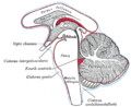
File:Gray768.png|Duret haemorrhages | |||
</gallery> | |||
Revision as of 01:20, 20 February 2025
Duret hemorrhages are small linear areas of bleeding in the midbrain and upper pons of the brainstem. These hemorrhages are typically associated with traumatic brain injury and are considered a secondary brainstem injury. They are named after the French neurologist Henri Duret, who first described them.
Pathophysiology
Duret hemorrhages occur due to the downward displacement of the brainstem, often as a result of increased intracranial pressure and brain herniation. The most common type of herniation associated with Duret hemorrhages is transtentorial herniation, where the temporal lobe is pushed downwards through the tentorial notch. This displacement causes stretching and tearing of the small penetrating arteries that supply the brainstem, leading to hemorrhage.
Clinical Significance
The presence of Duret hemorrhages is a grave prognostic sign, often indicating severe brain injury and poor outcome. They are typically seen in the context of severe head trauma or intracerebral hemorrhage that leads to significant brain swelling and herniation.
Diagnosis
Duret hemorrhages can be identified using neuroimaging techniques such as CT scan or MRI. On imaging, they appear as small, linear areas of hyperdensity (on CT) or hyperintensity (on MRI) in the midbrain and pons.
Treatment
There is no specific treatment for Duret hemorrhages themselves. Management focuses on addressing the underlying cause of increased intracranial pressure and preventing further brain herniation. This may involve surgical interventions such as decompressive craniectomy or medical management to reduce brain swelling.
Related Pages
-
 Duret haemorrhages

