Acoustic angiography: Difference between revisions
CSV import |
CSV import |
||
| Line 51: | Line 51: | ||
[[Category:Medical imaging]] | [[Category:Medical imaging]] | ||
[[Category:Ultrasound]] | [[Category:Ultrasound]] | ||
<gallery> | |||
File:Dual_element_transducer.png|Acoustic_angiography | |||
File:Wobbler_vs_Linear_Scanning_Configurations.png|Acoustic_angiography | |||
</gallery> | |||
Latest revision as of 01:11, 18 February 2025
Acoustic Angiography
Acoustic angiography is an advanced ultrasound imaging technique used to visualize the vascular system with high resolution and specificity. This method leverages the unique properties of microbubbles as contrast agents to enhance the imaging of blood vessels.
Principles[edit]
Acoustic angiography utilizes the nonlinear response of microbubbles to ultrasound waves. When exposed to ultrasound, these microbubbles oscillate and produce harmonic signals that can be detected and used to create detailed images of the vasculature. This technique is particularly useful for imaging small vessels and detecting abnormalities in the vascular structure.
Technology[edit]
The key component of acoustic angiography is the dual-element transducer, which consists of separate elements for transmitting and receiving ultrasound signals. This configuration allows for the isolation of harmonic signals generated by the microbubbles, enhancing the contrast and resolution of the images.
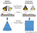
Scanning Configurations[edit]
There are different scanning configurations used in acoustic angiography, including wobbler and linear scanning.

- Wobbler Scanning: In this configuration, the transducer is mechanically wobbled to cover a larger area. This method is beneficial for imaging larger regions but may have limitations in terms of speed and resolution.
- Linear Scanning: This configuration involves moving the transducer linearly across the area of interest. It provides high-resolution images and is suitable for detailed studies of specific vascular regions.
Applications[edit]
Acoustic angiography is used in various medical fields, including:
- Cardiology: For imaging coronary arteries and detecting blockages or abnormalities.
- Oncology: To visualize tumor vasculature and assess the effectiveness of anti-angiogenic therapies.
- Neurology: For imaging cerebral vessels and diagnosing conditions such as aneurysms or arteriovenous malformations.
Advantages[edit]
The main advantages of acoustic angiography include:
- High spatial resolution, allowing for detailed visualization of small vessels.
- Non-invasive nature, reducing the risk associated with traditional angiography methods.
- Enhanced contrast due to the use of microbubbles, improving the detection of vascular abnormalities.
Limitations[edit]
Despite its advantages, acoustic angiography has some limitations:
- Limited penetration depth, which may restrict its use in imaging deep-seated vessels.
- Dependence on the availability and stability of microbubble contrast agents.
Future Directions[edit]
Research is ongoing to improve the technology and expand its clinical applications. Innovations in transducer design and microbubble formulation are expected to enhance the capabilities of acoustic angiography.
Related Pages[edit]
-
 Acoustic_angiography
 -
 Acoustic_angiography

