Ultramicrotomy: Difference between revisions
CSV import |
CSV import |
||
| Line 1: | Line 1: | ||
Ultramicrotomy | == Ultramicrotomy == | ||
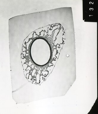
[[File:Salvinia cucullata megaspore.tif|thumb|right|Micrograph of a Salvinia cucullata megaspore prepared using ultramicrotomy.]] | |||
Ultramicrotomy | [[File:Cryo Ultramicrotome.jpg|thumb|right|A cryo ultramicrotome used for preparing samples at low temperatures.]] | ||
'''Ultramicrotomy''' is a technique used in the preparation of extremely thin sections of material for [[microscopy]], particularly [[transmission electron microscopy]] (TEM). This method is essential for examining the fine details of biological and material specimens at the [[nanometer]] scale. | |||
== Technique == | == Technique == | ||
Ultramicrotomy involves the use of an ultramicrotome, a specialized instrument designed to cut sections that are typically less than 100 nanometers thick. The process begins with embedding the specimen in a suitable medium, such as [[epoxy resin]], to provide support during sectioning. Once embedded, the sample is mounted on a specimen holder and trimmed to a suitable size. | |||
The ultramicrotome uses a diamond or glass knife to slice the specimen into thin sections. The knife is mounted on a vibrating arm, which moves back and forth to cut the sample. The sections are floated onto a water surface and then collected onto a grid for examination under a microscope. | |||
== Applications == | == Applications == | ||
== | Ultramicrotomy is widely used in the fields of [[biology]], [[materials science]], and [[nanotechnology]]. In biology, it allows for the detailed study of cellular structures, such as [[organelles]] and [[membranes]]. In materials science, it is used to analyze the microstructure of [[polymers]], [[metals]], and [[composites]]. | ||
* [[ | |||
* [[ | == Cryo-ultramicrotomy == | ||
* [[ | |||
Cryo-ultramicrotomy is a variation of the technique where the specimen is sectioned at cryogenic temperatures. This method is particularly useful for preserving the native state of biological samples, as it minimizes the damage caused by dehydration and chemical fixation. Cryo-ultramicrotomy is often used in conjunction with [[cryo-electron microscopy]] to study the structure of [[proteins]] and [[viruses]]. | |||
== Related pages == | |||
* [[Microscopy]] | |||
* [[Transmission electron microscopy]] | |||
* [[Cryo-electron microscopy]] | |||
* [[Microtome]] | |||
== References == | == References == | ||
* Glauert, A. M., & Lewis, P. R. (1998). ''Biological Specimen Preparation for Transmission Electron Microscopy''. Princeton University Press. | |||
* Bozzola, J. J., & Russell, L. D. (1999). ''Electron Microscopy: Principles and Techniques for Biologists''. Jones & Bartlett Learning. | |||
[[Category:Microscopy]] | [[Category:Microscopy]] | ||
[[Category:Scientific techniques]] | [[Category:Scientific techniques]] | ||
Revision as of 20:58, 9 February 2025
Ultramicrotomy

Ultramicrotomy is a technique used in the preparation of extremely thin sections of material for microscopy, particularly transmission electron microscopy (TEM). This method is essential for examining the fine details of biological and material specimens at the nanometer scale.
Technique
Ultramicrotomy involves the use of an ultramicrotome, a specialized instrument designed to cut sections that are typically less than 100 nanometers thick. The process begins with embedding the specimen in a suitable medium, such as epoxy resin, to provide support during sectioning. Once embedded, the sample is mounted on a specimen holder and trimmed to a suitable size.
The ultramicrotome uses a diamond or glass knife to slice the specimen into thin sections. The knife is mounted on a vibrating arm, which moves back and forth to cut the sample. The sections are floated onto a water surface and then collected onto a grid for examination under a microscope.
Applications
Ultramicrotomy is widely used in the fields of biology, materials science, and nanotechnology. In biology, it allows for the detailed study of cellular structures, such as organelles and membranes. In materials science, it is used to analyze the microstructure of polymers, metals, and composites.
Cryo-ultramicrotomy
Cryo-ultramicrotomy is a variation of the technique where the specimen is sectioned at cryogenic temperatures. This method is particularly useful for preserving the native state of biological samples, as it minimizes the damage caused by dehydration and chemical fixation. Cryo-ultramicrotomy is often used in conjunction with cryo-electron microscopy to study the structure of proteins and viruses.
Related pages
References
- Glauert, A. M., & Lewis, P. R. (1998). Biological Specimen Preparation for Transmission Electron Microscopy. Princeton University Press.
- Bozzola, J. J., & Russell, L. D. (1999). Electron Microscopy: Principles and Techniques for Biologists. Jones & Bartlett Learning.
