Duodenum: Difference between revisions
CSV import Tags: mobile edit mobile web edit |
CSV import |
||
| Line 1: | Line 1: | ||
= Duodenum = | |||
The '''duodenum''' is the first section of the [[small intestine]] in most higher vertebrates, including humans. It | The '''duodenum''' is the first section of the [[small intestine]] in most higher vertebrates, including humans. It is a hollow jointed tube connecting the [[stomach]] to the [[jejunum]]. | ||
==Anatomy== | == Anatomy == | ||
[[File:Tractus_intestinalis_duodenum.svg|thumb|right|Diagram of the duodenum and surrounding structures.]] | |||
The duodenum is approximately 25–30 cm (10–12 inches) long and is shaped like a "C". It is divided into four parts: | |||
The | |||
# The superior part | |||
The | # The descending part | ||
# The horizontal part | |||
# The ascending part | |||
The duodenum is located in the upper abdomen and is mostly retroperitoneal, meaning it is located behind the [[peritoneum]]. | |||
The | |||
== | == Histology == | ||
[[File:Dogduodenum100x3.jpg|thumb|left|Histological section of the duodenum showing the villi.]] | |||
The duodenum | The duodenum has a unique histological structure that includes: | ||
* [[Villi]]: Finger-like projections that increase the surface area for absorption. | |||
* [[Microvilli]]: Microscopic projections on the surface of the villi. | |||
* [[Brunner's glands]]: Located in the submucosa, these glands secrete an alkaline mucus that helps neutralize gastric acid. | |||
[[File:Microvilli-Duodenum.JPG|thumb|right|Microvilli of the duodenum under a microscope.]] | |||
== | == Function == | ||
[[File: | |||
[[File: | The primary function of the duodenum is to receive the chyme from the stomach and continue the process of digestion. It also plays a crucial role in: | ||
* Neutralizing stomach acid | |||
* Mixing chyme with digestive juices from the [[pancreas]] and [[bile]] from the [[liver]] | |||
* Absorbing nutrients such as iron and calcium | |||
== Clinical Significance == | |||
[[File:Small_bowel_duodenum_with_amyloid_deposition_congo_red_10X.jpg|thumb|left|Duodenum with amyloid deposition.]] | |||
The duodenum can be affected by various diseases and conditions, including: | |||
* [[Duodenal ulcer]]: A common form of [[peptic ulcer disease]]. | |||
* [[Celiac disease]]: An autoimmune disorder affecting the small intestine. | |||
* [[Giardiasis]]: An infection caused by the parasite ''Giardia lamblia''. | |||
[[File:Giardiasis_duodenum_low.jpg|thumb|right|Duodenum affected by giardiasis.]] | |||
== Development == | |||
The duodenum develops from the foregut and midgut during embryogenesis. It is initially a solid cord of cells that later becomes a hollow tube. | |||
== Related Pages == | |||
* [[Small intestine]] | * [[Small intestine]] | ||
* [[ | * [[Jejunum]] | ||
* [[Ileum]] | |||
* [[Stomach]] | |||
* [[Pancreas]] | * [[Pancreas]] | ||
== Gallery == | |||
<gallery> | |||
File:Gray1058.png|Diagram showing the position of the duodenum in the digestive system. | |||
File:Blausen_0817_SmallIntestine_Anatomy.png|Anatomy of the small intestine, including the duodenum. | |||
File:Gray533.png|Illustration of the duodenum and its relation to surrounding organs. | |||
</gallery> | |||
[[Category:Digestive system]] | [[Category:Digestive system]] | ||
[[Category:Gastroenterology]] | [[Category:Gastroenterology]] | ||
Latest revision as of 14:18, 21 February 2025
Duodenum[edit]
The duodenum is the first section of the small intestine in most higher vertebrates, including humans. It is a hollow jointed tube connecting the stomach to the jejunum.
Anatomy[edit]

The duodenum is approximately 25–30 cm (10–12 inches) long and is shaped like a "C". It is divided into four parts:
- The superior part
- The descending part
- The horizontal part
- The ascending part
The duodenum is located in the upper abdomen and is mostly retroperitoneal, meaning it is located behind the peritoneum.
Histology[edit]
The duodenum has a unique histological structure that includes:
- Villi: Finger-like projections that increase the surface area for absorption.
- Microvilli: Microscopic projections on the surface of the villi.
- Brunner's glands: Located in the submucosa, these glands secrete an alkaline mucus that helps neutralize gastric acid.
Function[edit]
The primary function of the duodenum is to receive the chyme from the stomach and continue the process of digestion. It also plays a crucial role in:
- Neutralizing stomach acid
- Mixing chyme with digestive juices from the pancreas and bile from the liver
- Absorbing nutrients such as iron and calcium
Clinical Significance[edit]
The duodenum can be affected by various diseases and conditions, including:
- Duodenal ulcer: A common form of peptic ulcer disease.
- Celiac disease: An autoimmune disorder affecting the small intestine.
- Giardiasis: An infection caused by the parasite Giardia lamblia.
Development[edit]
The duodenum develops from the foregut and midgut during embryogenesis. It is initially a solid cord of cells that later becomes a hollow tube.
Related Pages[edit]
Gallery[edit]
-
 Diagram showing the position of the duodenum in the digestive system.
 -
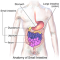 Anatomy of the small intestine, including the duodenum.
 -
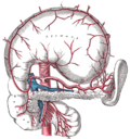 Illustration of the duodenum and its relation to surrounding organs.


